Far-Red Fluorescent FLICA® 660 Caspase-3/7 (DEVD) Assay Kit

This in vitro assay employs the far-red fluorescent inhibitor probe 660-DEVD-FMK to label active caspase 3 and caspase 7 enzymes in living cells. Analyze the fluorescent signal using fluorescence microscopy or flow cytometry.


SKU: 9125

Size: 25-50 Tests
Price:
Sale price$256.00

Our FLICA® probes are cell permeant, noncytotoxic Fluorescent Labeled Inhibitors of CAspases that covalently bind to active caspase enzymes. We have developed a far-red excitation and emission spectra FLICA® 660 probe for the detection of cells bearing active caspases 3 and 7.

Apoptosis is an evolutionarily conserved process of programmed cell death. It is centered on a cascade of proteolytic enzymes called caspases that are triggered in response to pro-apoptotic signals. Like the majority of other proteases, caspases are synthesized as pro-form precursors that undergo proteolytic maturation, either autocatalytically or in a cascade by enzymes with similar specificity. Active caspase enzymes consist of two large (~20 kD) and two small (~10 kD) subunits that non-covalently associate to form a two heterodimer, tetrameric active caspase. Once activated, caspases cleave protein substrates leading to the eventual disassembly of the cell. Caspases have been identified in organisms ranging from C. elegans to humans. Mammalian caspases play distinct roles in both apoptosis and inflammation.

Mammalian caspase enzymes have been classified as initiator, executioner, and inflammatory caspases. Once activated by initiator caspases (such as caspases -8, -9, and -10), executioner caspases -3 and/or -7 cleave specific sets of substrates that lead to apoptosis. Extrinsic activation of apoptosis, such as with FasL, TNF-a, or TRAIL binding to their associated death receptors, triggers the caspase-8 and -10-mediated cascade characteristic of the extrinsic apoptotic pathway, in which caspase-3 plays a dominant role. In intrinsic apoptosis activation, DNA damage or inhibition of DNA repair leads to cytochrome c release from mitochondria, triggering formation of the apoptosome and activation of caspase-9. Once active, caspase-9 cleaves pro-form precursors of effector caspases, such as -3 and -7, leading to the eventual disassembly of the cell.

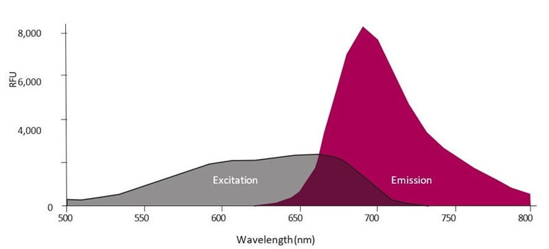
Our FLICA® 660 caspase-3/7 inhibitor probe contains the preferred binding sequence, Asp-Glu-Val-Asp (DEVD) for executioner caspases-3 and -7. This preferred caspase-3 and -7 binding sequence, DEVD, is labeled at the amino terminus end with a far-red fluorescent 660 dye and linked at the carboxyl end to a fluoromethyl ketone (FMK) reactive entity. The resulting cell permeant, fluorescent molecule, 660-DEVD-FMK, optimally excites at 660 nm and emits between 685-690 nm. A conventional red HeNe laser with a 633 nm excitation provides excellent excitation efficiency, enabling cells labeled with FLICA® 660 to be analyzed with most flow cytometers, as well as fluorescence microscopes equipped with electronic grey scale image capabilities.

To use FLICA®, add it directly to the cell media, incubate, and wash. FLICA® is cell-permeant and will efficiently diffuse in and out of all cells. If there is an active caspase-3 and/or -7 enzyme, it will covalently bind to FLICA® 660-DEVD-FMK and retain the far-red fluorescent signal within the cell. Unbound FLICA® will diffuse out of the cell during the wash steps. Apoptotic cells will retain a higher concentration of FLICA® and fluoresce brighter than non-apoptotic cells. There is no interference from pro-caspases or inactive forms of the enzyme. If the treatment is causing cell death via apoptosis, apoptotic cells will have an elevated level of caspase-3/7 activity relative to non-apoptotic or negative control cells and fluoresce with FLICA®. After labeling with FLICA®, cells can be counter-stained with other reagents and fixed or frozen.

660-DEVD-FMK
Caspase 3 and 7
660 nm / 690 nm
Flow cytometry, Fluorescence microscope
Cell culture
2-8°C
Domestic: Overnight Delivery; International: Priority Shipping
United States
  1. Prepare samples and controls
  2. Dilute 10X Apoptosis Wash Buffer 1:10 with diH2O.
  3. Reconstitute FLICA with 50 µL DMSO.
  4. Dilute FLICA 1:5 by adding 200 µL PBS.
  5. Add diluted FLICA to each sample at 1:30-1:60 (e.g. spike at 1:30 by adding 10 µL to 290 µL cultured cells).
  6. Incubate approximately 1 hour.
  7. Wash and spin cells three times.
  8. If desired, label with additional stains, such as Hoechst, DAPI, or an antibody.
  9. If desired, fix cells.
  10. Analyze with a fluorescence microscope or flow cytometer. FLICA 660 excites at 660 nm and emits at 680-690 nm.
  • FLICA Caspase-3/7 Inhibitor Reagent (660-DEVD-FMK), 1 vial, #6344
  • 10X Apoptosis Wash Buffer, 15 mL, #635
  • Fixative, 6 mL, #636
  • Kit Manual
  • Product Specific References

    PMID Publication
    37998361Spurlock, M., et al. 2023. The Inflammasome-Dependent Dysfunction and Death of Retinal Ganglion Cells after Repetitive Intraocular Pressure Spikes. Cells.
    35435672Li, Y, et al. 2022. Tumor Microenvironment-Responsive Yolk-Shell NaCl@Virus-Inspired Tetrasulfide-Organosilica for Ion-Interference Therapy via Osmolarity Surge and Oxidative Stress Amplification. ACS nano.
    35788117McLemore, AF, et al. 2022. Somatic gene mutations expose cytoplasmic DNA to co-opt the cGAS-STING-NLRP3 axis in Myelodysplastic syndromes. JCI insight.
    35967457Su, M, et al. 2022. Gasdermin D-dependent platelet pyroptosis exacerbates NET formation and inflammation in severe sepsis. Nature cardiovascular research, 732-747.
    36272266Deng, N, et al. 2022. Low-dose 5-fluorouracil ameliorates Th2 responses through the induction of apoptotic cell death of lung monocyte-derived dendritic cells in asthma. Biomedicine & pharmacotherapy, 113875.
    36373669Kodali, V, et al. 2022. Influence of Impurities from Manufacturing Process on the Toxicity Profile of Boron Nitride Nanotubes. Small (Weinheim an der Bergstrasse, Germany).

    Bulk Order Far-Red Fluorescent FLICA® 660 Caspase-3/7 (DEVD) Assay Kit

    Related Products

    Recently Viewed