Green Fluorescent FAM FLICA® Caspase-1 (WEHD) Assay Kit

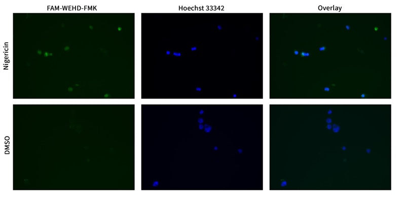
This in vitro assay employs the green fluorescent inhibitor probe FAM-WEHD-FMK to label active caspase-1 enzyme in living cells. Analyze samples using fluorescence microscopy, a fluorescence plate reader, or flow cytometry.


SKU: 9161

Size: 25 Tests
Price:
Sale price$234.00

Our FLICA® probes are non-cytotoxic Fluorescent Labeled Inhibitors of CAspases that covalently bind to active caspase enzymes. FLICA® measures the intracellular process of apoptosis instead of a side-effect, such as the turnover of phosphatidyl serine, and eliminates the incidence of false positives that often plagues methods like Annexin V and TUNEL staining. FLICA® can also be used to measure pyroptosis, a highly inflammatory form of programmed cell death.

To use FLICA®, add it directly to the cell culture media, incubate, and wash. FLICA® is cell-permeant and will efficiently diffuse in and out of all cells. If there is an active caspase enzyme inside the cell, it will covalently bind with FLICA® and retain the green fluorescent signal within the cell. Unbound FLICA® will diffuse out of the cell during the wash steps. Apoptotic and pyroptotic cells will retain a higher concentration of FLICA® and fluoresce brighter than healthy cells. There is no interference from pro-caspases or inactive forms of the enzymes. If the test treatment is causing cell death via apoptosis and/or pyroptosis, the cells will contain an elevated level of caspase activity relative to negative control cells and fluoresce with FLICA®.

Apoptosis is an evolutionarily conserved process of programmed cell suicide. It is centered on a cascade of proteolytic enzymes called caspases that are triggered in response to pro-apoptotic signals. Once activated, caspases cleave protein substrates leading to the eventual disassembly of the cell. Caspases have been identified in organisms ranging from C. elegans to humans. Mammalian caspases play distinct roles in both apoptosis and inflammation. In apoptosis, effector caspases (-3, -6, and -7) are responsible for proteolytic cleavages that lead to cell disassembly. Initiator caspases (-8, -9, and -10) regulate apoptosis upstream. Caspase-1 is associated with pyroptosis and inflammasome activity and takes on the role of a key housekeeping enzyme in its conversion of pro-IL-1ß protein into the active IL-1ß cytokine (Use FLICA® kits #98, #9122, and #9162 to detect caspase-1). Please note that macrophages and monocytes have been shown to rapidly secrete caspase-1 upon activation.

Like the majority of other proteases, caspases are synthesized as pro-form precursors that undergo proteolytic maturation, either autocatalytically or in a cascade by enzymes with similar specificity. Active caspase enzymes consist of two large (~20 kD) and two small (~10 kD) subunits that non-covalently associate to form a two heterodimer, tetrameric active caspase. Activated caspase enzymes cleave proteins by recognizing a 3 or 4 amino acid sequence that must include an aspartic acid (D) residue in the P1 position. This C–terminal residue is the target for the cleavage reaction at the carbonyl end. Each FLICA® probe contains a 3 or 4 amino acid sequence that is targeted by different activated caspases. This target sequence is sandwiched between a green fluorescent label, carboxyfluorescein (FAM), and a fluoromethyl ketone (FMK). Caspases cannot cleave the FLICA® inhibitor probe; instead, they form an irreversible covalent bond with the FMK on the target sequence and enzyme activity is inhibited. Our poly caspase FLICA® probe, FAM-VAD-FMK, can be used as a general reagent to detect apoptosis as it is recognized by many types of activated caspases. To more specifically target a particular caspase enzyme, use one of our specialized FLICA® reagents. We have kits for the detection of: caspase-1 (YVAD or WEHD) (also recognizes caspases 4 and 5), -2 (VDVAD), -3/7 (DEVD), -6 (VEID), -8 (LETD), -9 (LEHD), and -10 (AEVD). FLICA® kits are also available with a red or far red fluorescent label. Caspases, like most other crucial cell survival enzymes, are somewhat permissive in the target amino acid sequence they will recognize and cleave. Therefore, although FLICA® reagents contain the different amino acid target sequences preferred by each caspase, they can also recognize other active caspases when they are present. We encourage validation of caspase activity by an orthogonal technique.

FLICA® can be used to label suspension or adherent cells and thin tissue sections. After labeling with FAM-FLICA®, cells can be fixed or frozen. For tissues that will be paraffin-embedded after labeling, use our red sulforhodamine SR-FLICA® probes; do not use the green FAM-FLICA® probes as the FAM dye will be quenched during the paraffin embedding process.

Cells labeled with FAM-FLICA® can be counter-stained with reagents such as the red live/dead stains Propidium Iodide (included in FAM-FLICA® kits) and 7-AAD (catalog # 6163) to distinguish apoptosis from necrosis. Nuclear morphology can be concurrently observed using Hoechst 33342, a blue DNA binding dye (included in FLICA® kits). Cells can be viewed directly through a fluorescence microscope, or the fluorescence intensity can be quantified using a flow cytometer or fluorescence plate reader. FAM-FLICA® optimally excites at 488-492 nm and has a peak emission at 515-535 nm.

FAM-WEHD-FMK
Caspase-1
492 nm / 520 nm
Flow Cytometer, Fluorescence Microscope, Fluorescence Plate Reader
Cell culture
2-8°C
Domestic: Overnight Delivery; International: Priority Shipping
United States
  1. Prepare samples and controls
  2. Dilute 10X Apoptosis Wash Buffer 1:10 with diH20.
  3. Reconstitute FLICA with 50 μL DMSO.
  4. Dilute FLICA 1:5 by adding 200 μL PBS.
  5. Add diluted FLICA to each sample at 1:30 (e.g., add 10 μL to 290 μL of cultured cells).
  6. Incubate approximately 1 hour.
  7. Remove media and wash cells 3 times: add 1X Apoptosis Wash Buffer and spin cells.
  8. If desired, label with additional stains, such as Hoechst, Propidium Iodide, 7-AAD, or an antibody.
  9. If desired, fix cells.
  10. Analyze with a fluorescence microscope, fluorescence plate reader, or flow cytometer. FAM-FLICA excites at 492 nm and emits at 520 nm.

If working with adherent cells, please see the manual for additional protocols.

Kit 9161: 25 Tests
  • FLICA Caspase-1 Reagent (FAM-WEHD-FMK), 1 vial, #6708
  • 10X Apoptosis Wash Buffer, 15 mL, #635
  • Fixative, 6 mL, #636
  • Propidium Iodide, 1 mL, #638
  • Kit Manual
  • Kit 9162: 100 Tests
  • FLICA Caspase-1 Reagent (FAM-WEHD-FMK), 4 vials, #6708
  • 10X Apoptosis Wash Buffer, 60 mL, #634
  • Fixative, 6 mL, #636
  • Propidium Iodide, 1 mL, #638
  • Kit Manual
  • Product Specific References

    PMID Publication
    37121693Zhang, Y., et al. 2023. Blocking CXC Motif Chemokine Ligand 2 Ameliorates Diabetic Peripheral Neuropathy via Inhibiting Apoptosis and NLRP3 Inflammasome Activation. Biological & pharmaceutical bulletin, 672-683.

    Question: What is the difference between YVAD and WEHD and when is it an advantage using one or the other?

    Answer: Our new product offering FAM-WEHD-FMK is similar to our existing FAM-YVAD-FMK Assay. Both of these peptide sequences are known to target caspase 1,4, and 5. The WEHD sequence is thought to be a “better” caspase-1 target, as the kcat/kM rate is higher for WEHD vs YVAD (meaning faster conversion of substrate product by the enzyme). However, please note that if our understanding of how FLICA works is correct, the FLICA probe never actually binds to the enzyme via the YVAD or WEHD sequence, but rather the FMK moiety, then perhaps these faster binding kinetics are something of a moot point. In practice the performance characteristics of the two product are very similar. In our lab they were shown to be virtually indistinguishable. Nevertheless, we decided to carry both options so that customers can select their preferred targeting sequence based on their individual needs and experience.

    Question: Customer is not seeing a difference between control and induced cells(induction with LPS+ATP). Can we help with optimization? Parameters: macrophages induced from THP-1 cells, using 50 ng/ml PMA for 48 hr Cells in 12 well plates at 3×10^5 cells/well Three groups: experimental with HIV, Positive Control and Untreated. Given fresh media 24 hrs then added 1 ug/ml LPS for 24 hr then 5 mM ATP for 2 hr

    Answer: In our lab, we actually saw a greater response in the THP-1 monocytes (not PMA-primed), we had the greatest response with LPS exposure at 100 ng/mL + 5 mM ATP for 24 hours. In our THP I monocyte studies we found induction levels ranging from 10-30% (average was 26.2%) in 24 hour (LPS/ATP exposure) samples compared to 3-8% in negative controls. When working with THP-1 cells primed with PMA to become macrophage-like, in general we were able to achieve better results with lower LPS concentrations and exposure periods than with the THP-1 monocytes. For instance, exposure to 10ng/mL LPS for 2 hours without any supplemental ATP was sufficient to produce the desired effect. I am a bit concerned that the customer’s use of 1 ug/mL LPS for 24 hours may be too high concentration/exposure period and the susceptible cells are moving through pyroptosis, lysing, and are lost from the positive control sample well prior to even receiving the FAM-YVAD-FMK stain. If this is the case, they are missing the period when more of the positive control cells would be stain positive with FAM-FLICA. I would encourage them to experiment with lower LPS concentrations and exposure periods and see if their results are improved. It is also important to note caspase-1 is rapidly secreted by macrophages after its activation by the inflammasome pathway. Therefore, it turns out macrophages might not be the best cell model for use with this product. We have also been working with nigericin, as an alternate inducing agent.

    Question: The component FAM-YVAD-FMK Part#665 vial in the kit is empty. Please help me to solve this problem.

    Answer: All of our FLICA products, including FAM-YVAD-FMK, are lyophilized as part of the manufacturing process. The vials contain such a small amount of material (µg quantities) that the green FAM-FLICA reagents are nearly invisible in the amber vials. It may be visible as a slight iridescent sheen on the sides of the vial. Per the instructions in our manual, the FLICA vials are reconstituted in DMSO and diluted into PBS and subsequently diluted into cell culture media for staining cells. In order to check that the FLICA vial contains the proper lyophilized reagent, please check the appearance of the DMSO-reconstituted FLICA reagent. It should be orange in appearance and once diluted 1:5 in PBS, the FAM-FLICA reagent should be yellow in appearance.

    Related Products

    Recently Viewed