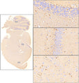
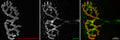
Anti-S100B Antibody
Our Anti-S100B chicken polyclonal primary antibody from Aves Labs is produced in-house. It detects human, mouse, and rat S100B, and is affinity-purified IgY. Works in ICC, WB.
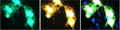
Immunofluorescent staining of COS-7 cells expressing S100B-flag using Aves Labs anti-S100B antibody (green) and rabbit anti-flag (red). DAPI nuclear stain (blue) shows nuclei of both transfected and untransfected cells. Staining shows 100% correspondence between chicken anti-S100B signal and anti-flag in transfected cells.
Click on image to zoom
SKU: S100B-0100
Ships: 1-2 business days
Product Details
S100B
S100 protein is a type of low molecular weight protein found in vertebrates characterized by two calcium binding sites of the helix-loop-helix ("EF-hand type") conformation. There are at least 21 different types of S100 proteins. The name is derived from the fact that the protein is 100% soluble in ammonium sulfate at neutral pH. Most S100 proteins are homodimeric, consisting of two identical polypeptides held together by non-covalent bonds. S100A is composed of an alpha and a beta chain whereas S100B is composed of two beta chains. Although S100 proteins are structurally similar to calmodulin, they differ in that they are cell-specific, expressed in particular cells at different levels depending on environmental factors. By contrast, calmodulin is a ubiquitous and universal intracellular Ca++ receptor widely expressed in many cells. S-100 is normally present in cells derived from the neural crest (Schwann cells, melanocytes, and glia cells), chondrocyte, adipocytes, myoepithelial cells, macrophages, Langerhans cells, dendritic cells, and keratinocytes. It may be present in some breast epithelial cells. This antibody stains schwannoma, ependymomas, astrogliomas, almost all benign and malignant melanoma and their metastasis.
Affinity-Purified IgY
0.2 mg/ml
Polyclonal
IgY
ICC, IHC, WB
Chicken
S100B
14 kDa
His tagged full length protein
Human
Human, Mouse, Rat
AB_2943661
Store at 4°C in the dark. Under these conditions, the antibodies should have a shelf life of at least twelve months, provided they remain sterile. For longer term storage, aliquot and freeze to avoid freeze-thaw of the antibody.
Liquid
Eggs from hens hyperimmunized with target were used to prepare an IgY fraction which was then subjected to antigen-specific affinity purification.
Phosphate-buffered (10 mM) isotonic (0.9%, w/v) saline (“PBS," pH 7.2) with sodium azide (0.02%, w/v) added as a preservative.
WB: 1:500-1:1000
IHC: 1:500-1:1000
ICC: 1:100-1:500
IHC: 1:500-1:1000
ICC: 1:100-1:500
No cross-reactivity reported.
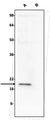
Each new lot of this antibody is tested in WB to confirm that it recognizes a single immunoreactive band of expected molecular weight when used to probe brain lysate.
These antibodies are to be used as research laboratory reagents and are not for use as diagnostic or therapeutic reagents in humans.
United States
12 months from receipt of product
NEF; S100 Calcium Binding Protein B; S100-B; Neural; S100beta; protein S100-B; S-100 calcium-binding protein; beta chain; S-100 protein subunit beta; S100 calcium-binding protein; beta (neural; S100 Calcium-Binding Protein B; S-100 Protein Beta Chain; S100 Calcium Binding Protein; S100; Beta
P04271
UniProt Summary: Small zinc- and- and calcium-binding protein that is highly expressed in astrocytes and constitutes one of the most abundant soluble proteins in brain.
Weakly binds calcium but binds zinc very tightly-distinct binding sites with different affinities exist for both ions on each monomer.
Physiological concentrations of potassium ion antagonize the binding of both divalent cations, especially affecting high-affinity calcium-binding sites.
Acts as a neurotrophic factor that promotes astrocytosis and axonal proliferation.
Involved in innervation of thermogenic adipose tissue by acting as an adipocyte-derived neurotrophic factor that promotes sympathetic innervation of adipose tissue.
Binds to and initiates the activation of STK38 by releasing autoinhibitory intramolecular interactions within the kinase.
Interaction with AGER after myocardial infarction may play a role in myocyte apoptosis by activating ERK1/2 and p53/TP53 signaling.
Could assist ATAD3A cytoplasmic processing, preventing aggregation and favoring mitochondrial localization.
May mediate calcium-dependent regulation on many physiological processes by interacting with other proteins, such as TPR-containing proteins, and modulating their activity
UniProt Summary: Small zinc- and- and calcium-binding protein that is highly expressed in astrocytes and constitutes one of the most abundant soluble proteins in brain.
Weakly binds calcium but binds zinc very tightly-distinct binding sites with different affinities exist for both ions on each monomer.
Physiological concentrations of potassium ion antagonize the binding of both divalent cations, especially affecting high-affinity calcium-binding sites.
Acts as a neurotrophic factor that promotes astrocytosis and axonal proliferation.
Involved in innervation of thermogenic adipose tissue by acting as an adipocyte-derived neurotrophic factor that promotes sympathetic innervation of adipose tissue.
Binds to and initiates the activation of STK38 by releasing autoinhibitory intramolecular interactions within the kinase.
Interaction with AGER after myocardial infarction may play a role in myocyte apoptosis by activating ERK1/2 and p53/TP53 signaling.
Could assist ATAD3A cytoplasmic processing, preventing aggregation and favoring mitochondrial localization.
May mediate calcium-dependent regulation on many physiological processes by interacting with other proteins, such as TPR-containing proteins, and modulating their activity
6285
Ice packs