Anti-Ki-67 Antibody
This antibody is the same product as PhosphoSolutions' catalog number 1006-Ki67, which has been discontinued.
Our Anti-Ki-67 primary antibody from Aves Labs is chicken polyclonal. It detects human, mouse, and rat Ki-67 and is total IgY fraction. It is great for use in WB, IHC, ICC.
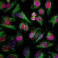
Immunostaining of HeLa cells showing specific labeling of Ki-67 (Cat. Ki67-0100, 1:2000, red) present in cytoplasmic microtubules. Additional immunostaining done with β-tubulin in green and nuclear staining with DAPI (blue). During cell cycle Ki-67 protein is predominantly expressed in the nucleoli of cells during mitosis and interphase, and is not present during quiescence.
Click on image to zoom
SKU: Ki67-1010
Ships: 1-2 business days
Product Details
Ki-67
Ki-67 is a nuclear protein that is considered to be essential for cellular proliferation since it is expressed only in active phases of the cell cycle (G1, S, G2 and mitosis) but not in resting phase G0 (Scholzen & Gerdes 2000). Ki-67 antibodies have therefore been widely used as markers of cell growth. Ki-67 has further been identified as a predicative and prognostic marker in breast, lung, cervical and other cancers (Yerushalmi et al., 2010; Folescu et al., 2018; Das et al., 2018).
Total IgY fraction
Polyclonal
IgY
ICC, IHC, WB
Chicken
MKI67
345-395 kDa
Recombinant human ki-67 protein expressed in and purified from E. Coli.
Human
Human, Mouse, Rat
AB_2941673
Recommended that the undiluted antibody be aliquoted into smaller working volumes (10-30 uL/vial depending on usage) upon arrival and stored long term at -20° C or -80° C, while keeping a working aliquot stored at 4° C for short term. Avoid freeze/thaw cycles. Stable for at least 1 year.
Liquid
Total IgY fraction
PBS + 10 mM NaN3
WB: 1:5000
IHC: 1:1000-1:5000
ICC: 1:1000-1:5000
IHC: 1:1000-1:5000
ICC: 1:1000-1:5000
Unconjugated
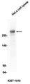
Specific for endogenous levels of the ~345 & 395 kDa Ki-67 isoforms. Additional bands are thought to be breakdown products of these 2 isoforms.
Western blots performed on each lot.
These antibodies are to be used as research laboratory reagents and are not for use as diagnostic or therapeutic reagents in humans.
After date of receipt, stable for at least 1 year at -20°C.
Antigen identified by monoclonal Ki 67; Antigen identified by monoclonal Ki-67; Antigen KI-67; Antigen KI67; Ki67; KI67_HUMAN; KIA; Marker of proliferation Ki-67; MIB 1; MIB; MKI67; PPP1R105; Proliferation marker protein Ki-67; Proliferation related Ki 67 antigen; Protein phosphatase 1 regulatory subunit 105; RP11-380J17.2
P46013
UniProt Summary: Protein that associates with the surface of mitotic chromosomes and acts both as a chromosome repellent during early mitosis and chromosome attractant during late mitosis. Required to maintain individual mitotic chromosomes dispersed in the cytoplasm following nuclear envelope disassembly. During early mitosis, relocalizes from nucleoli to the chromosome surface where it forms extended brush structures that cover a substantial fraction of the chromosome surface. The MKI67 brush structure prevents chromosomes from collapsing into a single chromatin mass by forming a steric and electrostatic charge barrier: the protein has a high net electrical charge and acts as a surfactant, dispersing chromosomes and enabling independent chromosome motility. During mitotic anaphase, the MKI67 brush structure collapses and MKI67 switches from a chromosome repellent to a chromosome attractant to promote chromosome clustering and facilitate the exclusion of large cytoplasmic particles from the future nuclear space. Mechanistically, dephosphorylation during mitotic exit and simultaneous exposure of a conserved basic patch induce the RNA-dependent formation of a liquid-like condensed phase on the chromosome surface, promoting coalescence of neighboring chromosome surfaces and clustering of chromosomes. Binds premature ribosomal RNAs during anaphase; promoting liquid-liquid phase separation. Binds DNA, with a preference for supercoiled DNA and AT-rich DNA. Does not contribute to the internal structure of mitotic chromosomes. May play a role in chromatin organization; it is however unclear whether it plays a direct role in chromatin organization or whether it is an indirect consequence of its function in mitotic chromosome.
4288
Blue Ice
General References
- Scholzen, T. and Gerdes, J., 2000. The Ki‐67 protein: from the known and the unknown. Journal of Cellular Physiology, 182(3), pp.311-322. PMID: 10653597
- Yerushalmi, R., Woods, R., Ravdin, P.M., Hayes, M.M. and Gelmon, K.A., 2010. Ki67 in breast cancer: prognostic and predictive potential. The Lancet Oncology, 11(2), pp.174-183. PMID: 20152769
- Folescu, R., Levai, C.M., GRIGORAŞ, M.L., Arghirescu, T.S., TALPOŞ, I.C., Gindac, C.M., ZAMFIR, C.L., Poroch, V. and Anghel, M.D., 2018. Expression and significance of Ki-67 in lung cancer. Romanian Journal of Morphology and Embryology, 59(1), pp.227-233. PMID: 29940632
- Das, D., Sengupta, M., Basu, K., Tirkey, M., Datta, C. and Chatterjee, U., 2018. Role of p16/Ki-67 Dual immunostaining in detection of cervical cancer precursors. Journal of Cytology, 35(3), p.153-158. PMID: 30089944