Ships: 1-2 business days
- Dilute Antigen Coating Buffer, 5X 1:5 using deionized water and mix for 15 minutes. As Antigen Coating Buffer is a 5X concentrate, crystalline precipitates may form in the bottle, especially when refrigerated. If this happens, gently warm the buffer until all crystals are dissolved. Do not let it boil.
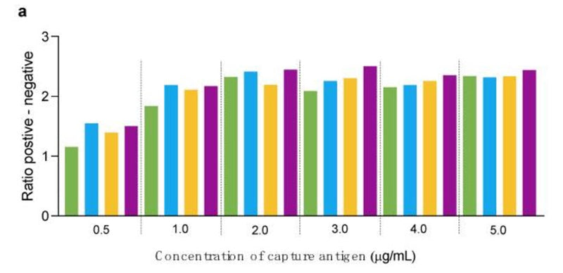
- Dilute your antigen into the coating buffer. Optimal coating concentration varies significantly from 0.2 µg/mL to 10 µg/mL.
- Let the solution stir 10 – 15 minutes and pipette onto the plate. Optimal coating volume generally ranges from 50 – 200 µL per well. ICT recommends coating antibodies onto Immulon II HB plates.
- Once added to the plate, incubate the coating solution from 8 – 24 hours at room temperature protected from light. Minimize evaporation by individually covering each plate with a plate sealing cover, wrapping a stack in plastic wrap, or placing plates in a humidified storage box and covering.
- After incubation, dump or aspirate the coating solution out of the wells.
- Wash the plate twice with ICT’s ELISA Wash Buffer.
- Aspirate and pipette one of ICT’s Blocking Buffers onto the plate at a higher volume than the coating solution (300 – 400 µL per well).
- Once added to the plate, incubate the block buffer from 8 – 24 hours at room temperature protected from light. Minimize evaporation by individually covering each plate with a plate sealing cover, wrapping a stack in plastic wrap, or placing plates in a humidified storage box and covering.
- Aspirate the block buffer.
- The assay can be run at this point, or the plate can be dried and packaged for later use.
- Dry the plate by letting it sit in the fume hood covered with foil overnight, or dry in a drying chamber under vacuum from 3 – 6 hours at room temperature. When dry, seal the plate in an air-tight foil pouch with a desiccant and store at 2-8°C protected from light.
Product Specific References
| PMID | Publication |
| 40005572 | Hernández, J, et al. 2025. Evaluation of IgM, IgA, and IgG Antibody Responses Against PCV3 and PCV2 in Tissues of Aborted Fetuses from Late-Term Co-Infected Sows. Pathogens, . |
| 38003105 | Cordero-Ortiz, M., et al. 2023. Development of a Multispecies Double-Antigen Sandwich ELISA Using N and RBD Proteins to Detect Antibodies against SARS-CoV-2. Animals: an open access journal from MDPI. |


