Anti-Splicing factor 3B subunit 4 (SF3B4) Antibody (3A1)

Our Anti-Splicing factor 3B subunit 4 (SF3B4) mouse monoclonal primary antibody detects bovine, human, mouse, other mammals (predicted), pig, and rat Splicing factor 3B subunit 4 (SF3B4), and is affinity-purified. It is validated for use in FC, ICC, WB.
![Left: Analysis of SF3B4 expression in HeLa cells by Immunocytochemistry. Cells were stained with mouse antibody to splicing factor SF3B4 (red, 1:1,000), and co-stained with chicken antibody to vimentin (C-1409-50, green, 1:10,000). Blue: DAPI nuclear stain. The SF3B4 antibody reveals strong granular staining of the nuclei, while the vimentin antibody specifically labels cytoplasmic intermediate filaments. Right: Western blot analysis of cell lysates, cytosol- or nuclear-enriched fractions, using mouse antibody to splicing factor SF3B4 (green, 1:1,000). [1] protein standard, [2] NIH-3T3 cytosolic fraction, [3] NIH-3T3 nuclear fraction, [4] HeLa cytosolic and [5] HeLa nuclear fractions. A strong single band at 49 kDa represents the SF3B4 protein, which is expressed exclusively in the nuclei. The same blot was simultaneously probed with rabbit anti-GAPDH antibody (R-1701-100, red, 1:20,000, lanes 2-5). The 37 kDa band corresponds to the GAPDH protein, detected mainly in the cytosolic fractions of these cells.](http://www.antibodiesinc.com/cdn/shop/files/m-1576-100-ihc-wb_120x120.jpg?v=1759273287)

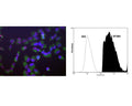
Left: Human HeLa cells stained with Mouse monoclonal antibody to splicing factor SF3B4 M-1576-100 (red), Chicken polyclonal antibody to Vimentin C-1409-50 (green) and DNA (blue, stained with DAPI). The monoclonal SF3B4 antibody reveals strong granular nuclear staining which is a little different from the DNA stain and presumably reflects splicosomal complexes. The polyclonal Vimentin antibody stains the cytoplasmic intermediate filament network of the HeLa cells. Right: Analysis of SF3B4 expression in rat pheochromocytoma PC-12 cell line by Flow Cytometry. Fixing and Permeabilization of cells: Absolute methanol (10 minutes in ice) and 0.1% Tween-20 in PBS, Blocking: 1% BSA, Primary antibody: Mouse Monoclonal antibody to SF3B4 (cat # M-1576-100, 2μg per ~10^6 cells) for 30 minutes at room temperature, Secondary antibody: Goat anti-mouse PE labeled secondary antibody (1:100 fold dilution) with incubation for 20 minutes in dark at room temperature. Non-specific Control IgG, clone X63 (cat # M-1249-100) was used as negative control under same conditions (black dashed). Flow cytometry data and results were generated using Orflo MoxiflowTM instrument and protocols.
Click on image to zoom
Ships: 1-2 business days