Anti-Parkinson disease protein 2 (Parkin) Antibody
Our Anti-Parkinson disease protein 2 (Parkin) rabbit polyclonal primary antibody detects human and rat Parkinson disease protein 2 (Parkin), and is whole serum. It is validated for use in IHC-Frozen, WB.
SKU: R-114-100
Ships: 1-2 business days
Product Details
Parkinson disease protein 2 (Parkin)
FUNCTION: Functions within a multiprotein E3 ubiquitin ligase complex, catalyzing the covalent attachment of ubiquitin moieties onto substrate proteins. These substrates include SYT11, CCNE1, GPR37, STUB1, a 22 kDa O-linked glycosylated isoform of SNCAIP and SEPT5. May play a more general role in the ubiquitin proteasomal pathway by participating in the removal and/or detoxification of abnormally folded or damaged protein. Loss of this ubiquitin ligase activity appears to be the mechanism underlying pathogenesis of PARK2. May protect neurons against alpha synuclein toxicity, proteasomal dysfunction, GPR37 accumulation, and kainate-induced excitotoxicity. May play a role in controlling neurotransmitter trafficking at the presynaptic terminal and in calcium-dependent exocytosis. Regulates cyclin E during neuronal apoptosis. May represent a tumor suppressor gene. SUBCELLULAR LOCATION: Cytoplasm. Co-localizes with STY11 in neutrites. Co-localizes with SNCAIP in brainstem Lewy bodies. TISSUE SPECIFICITY: Highly expressed in the brain including the substantia nigra. Expressed in heart, testis and skeletal muscle. Expression is down-regulated or absent in tumor biopsies, and absent in the brain of PARK2 patients. Overexpression protects dopamine neurons from kainate-mediated apoptosis.
Whole serum
Polyclonal
Mixed
IHC, WB
Rabbit
A synthetic peptide (NSLIKELHHFRILGEEQ) as part of human Parkin conjugated to KLH has been used as the immunogen.
Human
Human, Rat
Spin vial briefly before opening. Reconstitute in 100 µL sterile-filtered, ultrapure water. Centrifuge to remove any insoluble material. After reconstitution keep aliquots at -20°C for a higher stability, and at 2-8°C with an appropriate antibacterial agent. Glycerol (1:1) may be added for an additional stability. Avoid repetitive freeze/thaw cycles.
Lyophilized
WB: 1:2000
IHC: 1:1000
IHC: 1:1000
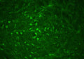
Nice staining is achieved in neuronal and cytoplasmic granules sections treated with citrate buffer for antigen retrieval. Few inclusions are stained but these were not positively identified as Lewy bodies.
Unconjugated
This antiserum is known to be highly specific for Parkin shown by IHC and WB. This antibody is known to react with rat and human Parkin.
For research use only.
United States
12 months after date of receipt (unopened vial).
AR JP; Ubiquitin E3 ligase PRKN; E3 ubiquitin ligase; Parkinson juvenile disease protein 2; E3 ubiquitin protein ligase parkin; Parkinson disease protein 2; E3 ubiquitin-protein ligase parkin; PARK2; FRA6E; PRKN; LPRS 2; LPRS2; PARK 2; Park2; Parkin 2; Parkinson disease (autosomal recessive juvenile 2; Parkinson disease (autosomal recessive juvenile 2 parkin; Parkinson disease protein 2; Parkinson juvenile disease protein 2; Parkinson protein 2 E3 ubiquitin protein ligase; Parkinson protein 2 E3 ubiquitin protein ligase (parkin; PDJ; PRKN 2; PRKN; PRKN2; PRKN2_HUMAN; Ubiquitin E3 ligase PRKN
O60260
UniProt Summary: Functions within a multiprotein E3 ubiquitin ligase complex, catalyzing the covalent attachment of ubiquitin moieties onto substrate proteins. Substrates include SYT11 and VDAC1. Other substrates are BCL2, CCNE1, GPR37, RHOT1/MIRO1, MFN1, MFN2, STUB1, SNCAIP, SEPTIN5, TOMM20, USP30, ZNF746, MIRO1 and AIMP2. Mediates monoubiquitination as well as 'Lys-6', 'Lys-11', 'Lys-48'-linked and 'Lys-63'-linked polyubiquitination of substrates depending on the context. Participates in the removal and/or detoxification of abnormally folded or damaged protein by mediating 'Lys-63'-linked polyubiquitination of misfolded proteins such as PARK7: 'Lys-63'-linked polyubiquitinated misfolded proteins are then recognized by HDAC6, leading to their recruitment to aggresomes, followed by degradation. Mediates 'Lys-63'-linked polyubiquitination of a 22 kDa O-linked glycosylated isoform of SNCAIP, possibly playing a role in Lewy-body formation. Mediates monoubiquitination of BCL2, thereby acting as a positive regulator of autophagy. Protects against mitochondrial dysfunction during cellular stress, by acting downstream of PINK1 to coordinate mitochondrial quality control mechanisms that remove and replace dysfunctional mitochondrial components. Depending on the severity of mitochondrial damage and/or dysfunction, activity ranges from preventing apoptosis and stimulating mitochondrial biogenesis to regulating mitochondrial dynamics and eliminating severely damaged mitochondria via mitophagy. Activation and recruitment onto the outer membrane of damaged/dysfunctional mitochondria (OMM) requires PINK1-mediated phosphorylation of both PRKN and ubiquitin. After mitochondrial damage, functions with PINK1 to mediate the decision between mitophagy or preventing apoptosis by inducing either the poly- or monoubiquitination of VDAC1, respectively; polyubiquitination of VDAC1 promotes mitophagy, while monoubiquitination of VDAC1 decreases mitochondrial calcium influx which ultimately inhibits apoptosis. When cellular stress results in irreversible mitochondrial damage, promotes the autophagic degradation of dysfunctional depolarized mitochondria (mitophagy) by promoting the ubiquitination of mitochondrial proteins such as TOMM20, RHOT1/MIRO1, MFN1 and USP30. Preferentially assembles 'Lys-6'-, 'Lys-11'- and 'Lys-63'-linked polyubiquitin chains, leading to mitophagy. The PINK1-PRKN pathway also promotes fission of damaged mitochondria by PINK1-mediated phosphorylation which promotes the PRKN-dependent degradation of mitochondrial proteins involved in fission such as MFN2. This prevents the refusion of unhealthy mitochondria with the mitochondrial network or initiates mitochondrial fragmentation facilitating their later engulfment by autophagosomes. Regulates motility of damaged mitochondria via the ubiquitination and subsequent degradation of MIRO1 and MIRO2; in motor neurons, this likely inhibits mitochondrial intracellular anterograde transport along the axons which probably increases the chance of the mitochondria undergoing mitophagy in the soma. Involved in mitochondrial biogenesis via the 'Lys-48'-linked polyubiquitination of transcriptional repressor ZNF746/PARIS which leads to its subsequent proteasomal degradation and allows activation of the transcription factor PPARGC1A. Limits the production of reactive oxygen species (ROS). Regulates cyclin-E during neuronal apoptosis. In collaboration with CHPF isoform 2, may enhance cell viability and protect cells from oxidative stress. Independently of its ubiquitin ligase activity, protects from apoptosis by the transcriptional repression of p53/TP53. May protect neurons against alpha synuclein toxicity, proteasomal dysfunction, GPR37 accumulation, and kainate-induced excitotoxicity. May play a role in controlling neurotransmitter trafficking at the presynaptic terminal and in calcium-dependent exocytosis. May represent a tumor suppressor gene.

5071
25°C (ambient)