Anti-ERK1 (C-terminal region) Antibody (M233)
Our ERK1 (C-terminal region) primary antibody from PhosphoSolutions is mouse monoclonal. It detects human, mouse, and rat ERK1 (C-terminal region) and is Protein A purified. It is great for use in WB, ICC, IHC.
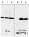
Western blot analysis of human A431 epithelial cells untreated (lanes 1 & 4) or treated with 100 nM calyculin A for 30 min. (lanes 2 & 5) or 100 ng/ml EGF for 60 min. (lanes 3 & 6). The blots were probed with anti-ERK1 (C-terminal region) (lanes 1, 2, & 3) or anti-ERK1/2 (Thr-202/Tyr-204) (lanes 4, 5, & 6).
Click on image to zoom
SKU: EM2331
Ships: 1-2 business days
Product Details
ERK1 (C-terminal region)
Mitogen-activated protein kinases (MAPKs) are a widely conserved family of serine/threonine protein kinases involved in many cellular programs such as cell proliferation, differentiation, motility, and death. The ERK1/2 (p44/42) signaling pathway can be activated in response to a diverse range of extracellular stimuli including mitogens, growth factors, and cytokines. Upon stimulation, a sequential three-part protein kinase cascade is initiated, consisting of a MAP kinase kinase kinase (MAPKKK), a MAP kinase kinase (MAPKK), and a MAP kinase (MAPK). Multiple ERK1/2 MAPKKKs have been identified, including members of the Raf family as well as Mos and Tpl2/Cot. MEK1 and MEK2 are the primary MAPKKs in this pathway. MEK1 and MEK2 activate ERK1 and ERK2 through phosphorylation of activation loop residues Thr-202/Tyr-204 and Thr-185/Tyr-187, respectively. ERK1/2 are negatively regulated by a family of dual-specificity (Thr/Tyr) MAPK phosphatases. Several downstream targets of ERK1/2 have been identified, including p90RSK and the transcription factor Elk-1.
Protein A Purified
Monoclonal
M233
IgG1
ELISA, ICC, IHC, WB
Mouse
MAPK1
44
Clone (M233) was generated from a recombinant human ERK1 protein that included amino acids residues in the C-terminal region. This sequence is conserved in rat and mouse ERK1, and has low homology to ERK2, as well as other ERK family members.
Human
Human, Mouse, Rat
Storage at -20°C is recommended, as aliquots may be taken without freeze/thawing due to presence of 50% glycerol. Stable for at least 1 year at -20°C.
Liquid
PBS + 1 mg/ml BSA, 0.05% NaN3 and 50% glycerol
WB: 1:1000
ICC: 1:100
IP: 1:100
ICC: 1:100
IP: 1:100
Unconjugated
The antibody detects a 44 kDa* protein corresponding to ERK1 on SDS-PAGE immunoblots of human A431 epithelial cells. It does not detect ERK2.
Western blots performed on each lot.
For research use only. Not intended for therapeutic or diagnostic use. Use of all products is subject to our terms and conditions, which can be viewed on our website.
United States
After date of receipt, stable for at least 1 year at -20°C.
ERK 1; ERK; ERK 2; ERK-2; ERK1; erk1/2; ERK2; ERT1; ERT2; Extracellular signal regulated kinase 1; Extracellular signal-regulated kinase 2; MAP kinase 1; MAP kinase 2; MAP kinase isoform p42; MAP kinase isoform p44; MAPK 1; MAPK 2; MAPK 3; Mapk1; MAPK2; MAPK3; Mitogen-activated protein kinase 1; Mitogen-activated protein kinase 2; MK01_HUMAN; p38; p40; p41; p42-MAPK; PRKM 2; p42; p44; MAPK
P28482
UniProt Summary: Serine/threonine kinase which acts as an essential component of the MAP kinase signal transduction pathway. MAPK1/ERK2 and MAPK3/ERK1 are the 2 MAPKs which play an important role in the MAPK/ERK cascade. They participate also in a signaling cascade initiated by activated KIT and KITLG/SCF. Depending on the cellular context, the MAPK/ERK cascade mediates diverse biological functions such as cell growth, adhesion, survival and differentiation through the regulation of transcription, translation, cytoskeletal rearrangements. The MAPK/ERK cascade also plays a role in initiation and regulation of meiosis, mitosis, and postmitotic functions in differentiated cells by phosphorylating a number of transcription factors. About 160 substrates have already been discovered for ERKs. Many of these substrates are localized in the nucleus, and seem to participate in the regulation of transcription upon stimulation. However, other substrates are found in the cytosol as well as in other cellular organelles, and those are responsible for processes such as translation, mitosis and apoptosis. Moreover, the MAPK/ERK cascade is also involved in the regulation of the endosomal dynamics, including lysosome processing and endosome cycling through the perinuclear recycling compartment (PNRC); as well as in the fragmentation of the Golgi apparatus during mitosis. The substrates include transcription factors (such as ATF2, BCL6, ELK1, ERF, FOS, HSF4 or SPZ1), cytoskeletal elements (such as CANX, CTTN, GJA1, MAP2, MAPT, PXN, SORBS3 or STMN1), regulators of apoptosis (such as BAD, BTG2, CASP9, DAPK1, IER3, MCL1 or PPARG), regulators of translation (such as EIF4EBP1 and FXR1) and a variety of other signaling-related molecules (like ARHGEF2, DCC, FRS2 or GRB10). Protein kinases (such as RAF1, RPS6KA1/RSK1, RPS6KA3/RSK2, RPS6KA2/RSK3, RPS6KA6/RSK4, SYK, MKNK1/MNK1, MKNK2/MNK2, RPS6KA5/MSK1, RPS6KA4/MSK2, MAPKAPK3 or MAPKAPK5) and phosphatases (such as DUSP1, DUSP4, DUSP6 or DUSP16) are other substrates which enable the propagation the MAPK/ERK signal to additional cytosolic and nuclear targets, thereby extending the specificity of the cascade. Mediates phosphorylation of TPR in response to EGF stimulation. May play a role in the spindle assembly checkpoint. Phosphorylates PML and promotes its interaction with PIN1, leading to PML degradation. Phosphorylates CDK2AP2. Phosphorylates phosphoglycerate kinase PGK1 under hypoxic conditions to promote its targeting to the mitochondrion and suppress the formation of acetyl-coenzyme A from pyruvate. Phosphorylates GJA1 at 'Ser-279' and 'Ser-282' resulting in an increase in GJA1 ubiquitination and ultimately lysosomal degradation.

5594
Blue Ice