Anti-RhoA (Ser-188), Phosphospecific Antibody
Our RhoA (Ser-188) rabbit polyclonal phosphospecific primary antibody from PhosphoSolutions is produced in-house. It detects human, mouse, and rat RhoA (Ser-188) and is antigen affinity purified. It is great for use in WB.
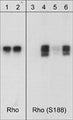
Western blot analysis of human RhoA GST fusion recombinant unphosphorylated (lanes 1 & 3) or phosphorylated with PKA (lanes 2, 4, 5 & 6). The blots were probed with anti-Rho (RP1501; lanes 1 & 2) or with anti-RhoA (Ser-188) (RP1361; lanes 3-6). The latter antibody was used in the presence of no peptide (lanes 3 & 4), phospho-Rho (Ser-188) peptide (lane 5), or a non-specific phosphoserine peptide (lane 6).
Click on image to zoom
SKU: RP1361
Ships: 1-2 business days
Product Details
RhoA (Ser-188)
Rho (A, B, & C) proteins are members of the Ras superfamily of GTPases. These proteins regulate a variety of cellular functions, including cell cycle progression, cytoskeletal rearrangement, and gene expression. Rho cycles between the active GTP-bound form and an inactive GDP-bound form. Interconversion between these forms is controlled by guanine nucleotide exchange factors (GEFs) and GTPase-activating proteins (GAPs). The Rho proteins RhoA, RhoB, and RhoC are highly homologous and contain the consensus amino acid sequences necessary for GDP/GTP-binding and GTPase activity. Post-translational regulation of Rho activity has been shown specifically for RhoA. This Rho protein is phosphorylated in vitro on serine 188 by cAMP- and cGMP-dependent kinases (PKA and PKG). Ser-188 phosphorylation enhances RhoGDI binding and inhibits RhoA-mediated stress fiber formation. Thus, Ser-188 is an important site for negative regulation of RhoA activity.
Antigen Affinity Purified
Polyclonal
IgG
ELISA, WB
Rabbit
RHOA
22
Phospho-RhoA (Ser-188) synthetic peptide (coupled to KLH) corresponding to amino acid residues around serine 188 of human RhoA. This peptide sequence is highly conserved in rat, mouse, and chicken RhoA, but is not found in RhoB or RhoC proteins.
Human
Human, Mouse, Rat
Storage at -20°C is recommended, as aliquots may be taken without freeze/thawing due to presence of 50% glycerol. Stable for at least 1 year at -20°C.
Liquid
PBS + 1 mg/ml BSA, 0.05% NaN3 and 50% glycerol
WB: 1:1000
Unconjugated
The antibody detects a RhoA GST fusion protein when phosphorylated with PKA, but does not detect the unphosphorylated recombinant protein.
Phosphorylated
Ser-188
Western blots performed on each lot.
For research use only. Not intended for therapeutic or diagnostic use. Use of all products is subject to our terms and conditions, which can be viewed on our website.
United States
After date of receipt, stable for at least 1 year at -20°C.
ARH12; RHO12; RHOH12; transforming protein RhoA; Aplysia ras-related homolog 12; oncogene RHO H12; small GTP binding protein RhoA
P06749
UniProt Summary: Function Small GTPase which cycles between an active GTP-bound and an inactive GDP-bound state. Mainly associated with cytoskeleton organization, in active state binds to a variety of effector proteins to regulate cellular responses such as cytoskeletal dynamics, cell migration and cell cycle.
Regulates a signal transduction pathway linking plasma membrane receptors to the assembly of focal adhesions and actin stress fibers.
Involved in a microtubule-dependent signal that is required for the myosin contractile ring formation during cell cycle cytokinesis.
Plays an essential role in cleavage furrow formation. Required for the apical junction formation of keratinocyte cell-cell adhesion.
Essential for the SPATA13-mediated regulation of cell migration and adhesion assembly and disassembly.
The MEMO1-RHOA-DIAPH1 signaling pathway plays an important role in ERBB2-dependent stabilization of microtubules at the cell cortex. It controls the localization of APC and CLASP2 to the cell membrane, via the regulation of GSK3B activity. In turn, membrane-bound APC allows the localization of the MACF1 to the cell membrane, which is required for microtubule capture and stabilization.
Involved in the reorientation of endothelial cells and their actin stress fibers in response to cellular mechantransduction-mediated activation by ARHGEF40.
Regulates KCNA2 potassium channel activity by reducing its location at the cell surface in response to CHRM1 activation; promotes KCNA2 endocytosis.
Acts as an allosteric activator of guanine nucleotide exchange factor ECT2 by binding in its activated GTP-bound form to the PH domain of ECT2 which stimulates the release of PH inhibition and promotes the binding of substrate RHOA to the ECT2 catalytic center.
May be an activator of PLCE1.
In neurons, involved in the inhibition of the initial spine growth. Upon activation by CaMKII, modulates dendritic spine structural plasticity by relaying CaMKII transient activation to synapse-specific, long-term signaling.
Acts as a regulator of platelet alpha-granule release during activation and aggregation of platelets.
When activated by DAAM1 may signal centrosome maturation and chromosomal segregation during cell division. May also be involved in contractile ring formation during cytokinesis

UniProt Summary: Function Small GTPase which cycles between an active GTP-bound and an inactive GDP-bound state. Mainly associated with cytoskeleton organization, in active state binds to a variety of effector proteins to regulate cellular responses such as cytoskeletal dynamics, cell migration and cell cycle.
Regulates a signal transduction pathway linking plasma membrane receptors to the assembly of focal adhesions and actin stress fibers.
Involved in a microtubule-dependent signal that is required for the myosin contractile ring formation during cell cycle cytokinesis.
Plays an essential role in cleavage furrow formation. Required for the apical junction formation of keratinocyte cell-cell adhesion.
Essential for the SPATA13-mediated regulation of cell migration and adhesion assembly and disassembly.
The MEMO1-RHOA-DIAPH1 signaling pathway plays an important role in ERBB2-dependent stabilization of microtubules at the cell cortex. It controls the localization of APC and CLASP2 to the cell membrane, via the regulation of GSK3B activity. In turn, membrane-bound APC allows the localization of the MACF1 to the cell membrane, which is required for microtubule capture and stabilization.
Involved in the reorientation of endothelial cells and their actin stress fibers in response to cellular mechantransduction-mediated activation by ARHGEF40.
Regulates KCNA2 potassium channel activity by reducing its location at the cell surface in response to CHRM1 activation; promotes KCNA2 endocytosis.
Acts as an allosteric activator of guanine nucleotide exchange factor ECT2 by binding in its activated GTP-bound form to the PH domain of ECT2 which stimulates the release of PH inhibition and promotes the binding of substrate RHOA to the ECT2 catalytic center.
May be an activator of PLCE1.
In neurons, involved in the inhibition of the initial spine growth. Upon activation by CaMKII, modulates dendritic spine structural plasticity by relaying CaMKII transient activation to synapse-specific, long-term signaling.
Acts as a regulator of platelet alpha-granule release during activation and aggregation of platelets.
When activated by DAAM1 may signal centrosome maturation and chromosomal segregation during cell division. May also be involved in contractile ring formation during cytokinesis
387
Blue Ice