Anti-Insulin Receptor (Thr1160) Antibody
Our Anti-Insulin Receptor (Thr1160) rabbit polyclonal phosphospecific primary antibody from PhosphoSolutions is produced in-house. It detects human Insulin Receptor (Thr1160) and is antigen affinity purified from pooled serum. It is great for use in WB.
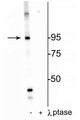
Western blot of HeLa cell lysate showing specific labeling of the ~95 kDa IR protein phosphorylated at Thr1160 in the first lane (-). Phosphospecificity is shown in the second lane (+) where immunolabeling is completely eliminated by blot treatment with lambda phosphatase (λ-Ptase, 1200 units for 30 min).
Click on image to zoom
SKU: p168-1160
Ships: 1-2 business days
Product Details
Insulin Receptor (Thr1160)
The insulin receptor (IR) is a well-studied receptor tyrosine kinase composed of two α subunits, responsible for the extracellular insulin binding site, and two β subunits, responsible for intracellular protein kinase activity (Endemann et al., 1990, Chiu et al., 2010). The binding of insulin to the α subunits activates the intrinsic kinase activity located in the β subunits and subsequently initiates a cascade of phosphorylation events causing major conformational changes in the activation loop of the kinase domain, which lead to different biological functions (Chiu et al., 2010). It has been hypothesized that Thr-1160 phosphorylation affects or is affected by Tyr-1158/62/63 phosphorylation and that the conformation of Thr-1160 and pThr-1160 is likely to be affected by the phosphorylation status of the surrounding tyrosines.
Antigen Affinity Purified from Pooled Serum
Polyclonal
IgG
ELISA, WB
Rabbit
INSR
95 kDa
Synthetic phospho-peptide corresponding to amino acid residues surrounding Thr1160 of human insulin receptor, conjugated to keyhole limpet hemocyanin (KLH).
Human
Human
Bovine, Canine, Non-Human Primate, Mouse, Rat, Xenopus
AB_2492137
Storage at -20°C is recommended, as aliquots may be taken without freeze/thawing due to presence of 50% glycerol. Stable for at least 1 year at -20°C.
Liquid
Prepared from pooled rabbit serum by affinity purification via sequential chromatography on phospho and non-phosphopeptide affinity columns.
10 mM HEPES (pH 7.5), 150 mM NaCl, 100 µg per ml BSA and 50% glycerol.
WB: 1:1000
Unconjugated
Specific for endogenous levels of the ~95 kDa Insulin receptor phosphorylated at Thr1160. Immunolabeling is completely eliminated with λ-phosphatase treatment.
Phosphorylated
Thr1160
Western blots performed on each lot.
For research use only. Not intended for therapeutic or diagnostic use. Use of all products is subject to our terms and conditions, which can be viewed on our website.
After date of receipt, stable for at least 1 year at -20°C.
CD220; HHF5; human insulin receptor; Insr; INSR_HUMAN; Insulin receptor subunit beta; IR 1; IR; IR-1; IR1 ,
P06213
UniProt Summary: Receptor tyrosine kinase which mediates the pleiotropic actions of insulin. Binding of insulin leads to phosphorylation of several intracellular substrates, including, insulin receptor substrates (IRS1, 2, 3, 4), SHC, GAB1, CBL and other signaling intermediates. Each of these phosphorylated proteins serve as docking proteins for other signaling proteins that contain Src-homology-2 domains (SH2 domain) that specifically recognize different phosphotyrosine residues, including the p85 regulatory subunit of PI3K and SHP2. Phosphorylation of IRSs proteins lead to the activation of two main signaling pathways: the PI3K-AKT/PKB pathway, which is responsible for most of the metabolic actions of insulin, and the Ras-MAPK pathway, which regulates expression of some genes and cooperates with the PI3K pathway to control cell growth and differentiation. Binding of the SH2 domains of PI3K to phosphotyrosines on IRS1 leads to the activation of PI3K and the generation of phosphatidylinositol-(3, 4, 5)-triphosphate (PIP3), a lipid second messenger, which activates several PIP3-dependent serine/threonine kinases, such as PDPK1 and subsequently AKT/PKB. The net effect of this pathway is to produce a translocation of the glucose transporter SLC2A4/GLUT4 from cytoplasmic vesicles to the cell membrane to facilitate glucose transport. Moreover, upon insulin stimulation, activated AKT/PKB is responsible for: anti-apoptotic effect of insulin by inducing phosphorylation of BAD; regulates the expression of gluconeogenic and lipogenic enzymes by controlling the activity of the winged helix or forkhead (FOX) class of transcription factors. Another pathway regulated by PI3K-AKT/PKB activation is mTORC1 signaling pathway which regulates cell growth and metabolism and integrates signals from insulin. AKT mediates insulin-stimulated protein synthesis by phosphorylating TSC2 thereby activating mTORC1 pathway. The Ras/RAF/MAP2K/MAPK pathway is mainly involved in mediating cell growth, survival and cellular differentiation of insulin. Phosphorylated IRS1 recruits GRB2/SOS complex, which triggers the activation of the Ras/RAF/MAP2K/MAPK pathway. In addition to binding insulin, the insulin receptor can bind insulin-like growth factors (IGFI and IGFII). Isoform Short has a higher affinity for IGFII binding. When present in a hybrid receptor with IGF1R, binds IGF1. PubMed:12138094 shows that hybrid receptors composed of IGF1R and INSR isoform Long are activated with a high affinity by IGF1, with low affinity by IGF2 and not significantly activated by insulin, and that hybrid receptors composed of IGF1R and INSR isoform Short are activated by IGF1, IGF2 and insulin. In contrast, PubMed:16831875 shows that hybrid receptors composed of IGF1R and INSR isoform Long and hybrid receptors composed of IGF1R and INSR isoform Short have similar binding characteristics, both bind IGF1 and have a low affinity for insulin. In adipocytes, inhibits lipolysis (By similarity). {ECO:0000250|UniProtKB:P15208, ECO:0000269|PubMed:12138094, ECO:0000269|PubMed:16314505, ECO:0000269|PubMed:16831875, ECO:0000269|PubMed:8257688, ECO:0000269|PubMed:8276809, ECO:0000269|PubMed:8452530, ECO:0000269|PubMed:9428692}.

3643
Blue Ice
- Western Blot Protocol: Download
General References
- Chiu SL and Cline HL (2010). Insulin receptor signaling in the development of neuronal structure and function. Neural Dev. 5:7. PMID: 17544402
- Endemann G, Yonezawa K, and Roth RA (1990). Phosphatidylinositol kinase or an associated protein is a substrate for the insulin receptor tyrosine kinase. J. Biol. Chem. 265:396-400. PMID: 16038406