Anti-E-Cadherin (Cytoplasmic) Antibody (M168)
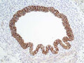
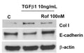
Our E-Cadherin (Cytoplasmic) mouse monoclonal primary antibody from PhosphoSolutions is produced in-house. It detects human, mouse, and rat E-Cadherin (Cytoplasmic) and is Protein A purified. It is great for use in WB, ICC, IHC, IP.
SKU: CM1681
Ships: 1-2 business days
Product Details
E-Cadherin (Cytoplasmic)
Cadherins are transmembrane glycoproteins vital in calcium-dependent cell-cell adhesion during tissue differentiation. Cadherins cluster to form foci of homophilic binding units. A key determinant to the strength of the cadherin-mediated adhesion may be by the juxtamembrane region in cadherins. This region induces clustering and also binds to the protein p120 catenin. The cytoplasmic region is highly conserved in sequence and has been shown experimentally to regulate the cell-cell binding function of the extracellular domain of E-cadherin, possibly through interaction with the cytoskeleton. Many cadherins are regulated by phosphorylation, including N-cadherin and E-cadherin. N-cadherin is phosphorylated by c-Src at Tyr-820, Tyr-853, Tyr-860, Tyr-884, and Tyr-886. Phosphorylation of Tyr-860 can disrupt cadherin binding to β-catenin. Since many of these tyrosine sites are conserved in the cadherin family, phosphorylation of these sites may be critical for cadherin function.
Protein A Purified
Monoclonal
M168
IgG1
ELISA, ICC, IHC, IP, WB
Mouse
CDH1
120
Clone (M168) was generated from a mouse recombinant E-Cadherin protein containing amino acids in the cytoplasmic region. This sequence is highly conserved in human and rat E-cadherin.
Mouse
Human, Mouse, Rat
Storage at -20°C is recommended, as aliquots may be taken without freeze/thawing due to presence of 50% glycerol. Stable for at least 1 year at -20°C.
Liquid
PBS + 1 mg/ml BSA, 0.05% NaN3 and 50% glycerol
WB: 1:1000
IHC: 1:50
ICC: 1:250
IP: 1:100
IHC: 1:50
ICC: 1:250
IP: 1:100
Unconjugated
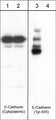
This E-cadherin antibody detects a 120 kDa* protein in human A431 cells, and does not cross-react with VE-cadherin or N-cadherin.
Western blots performed on each lot.
For research use only. Not intended for therapeutic or diagnostic use. Use of all products is subject to our terms and conditions, which can be viewed on our website.
United States
After date of receipt, stable for at least 1 year at -20°C.
AA960649; Uvomorulin; E-cad; L-CAM; E-cadherin; Cadherin-1; CTF1; CTF2; CTF3; CD324; Epithelial Cadherin
P09803
UniProt Summary: Cadherins are calcium-dependent cell adhesion proteins. They preferentially interact with themselves in a homophilic manner in connecting cells; cadherins may thus contribute to the sorting of heterogeneous cell types. CDH1 is involved in mechanisms regulating cell-cell adhesions, mobility and proliferation of epithelial cells. Promotes organization of radial actin fiber structure and cellular response to contractile forces, via its interaction with AMOTL2 which facilitates anchoring of radial actin fibers to CDH1 junction complexes at the cell membrane. Plays a role in the early stages of desmosome cell-cell junction formation via facilitating the recruitment of DSG2 and DSP to desmosome plaques. Has a potent invasive suppressor role. It is a ligand for integrin alpha-E/beta-7.
12550
Blue Ice