Our Anti-Integrin beta 2 alpha subunit mouse monoclonal primary antibody detects rat Integrin beta 2 alpha subunit, and is IgG. It is validated for use in FC, ICC, IHC-Paraffin-embedded, IP.
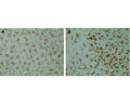
Immunohistochemical staining with mouse monoclonal antibody to CD11b [OX42] M-1325-100 of pure (>98%) microglial cells isolated from mixed glia cultures obtained from neonate rat brains (1.25 µg/mL). Secondary antibody was biotinylated anti-mouse (1.25 µg/mL) and detection method used Extravidin-Cy3 conjugate (2.5 µg/mL). Cells were fixed before incubation with OX42 (1hr). Cells were not permeabilized. Pictures courtesy of Markus Smolny.
Clone OX-42 recognizes the rat equivalent of human CD11b and shares a common epitope with CB11c (integrin apha M and alpha X chains). (PMID:1672643; Tamatani T et al 1991). CD11b is a single-pass type I membrane protein that belongs to the integrin alpha chain family. CD11b is predominantly expressed in monocytes and granulocytes and is implicated in various adhesive interactions of monocytes, macrophages and granulocytes as well as in mediating the uptake of complement-coated particles (Ref: SWISSPROT). CD11b is also frequently used as a microglial marker allowing to distinguish between quiescent and activated microglia based on the intensity of CD11b staining. Moreover the OX-42 monoclonal antibody specifically binds to the CR3 complement (C3bi) receptor found on most monocytes, granulocytes, macrophages, dendritic cells, and microglia. OX-42 antibody inhibits C3bi binding activity. CD11b, also known as integrin alpha M or Mac-1, and is a component of complement receptor 3 (CR3). CD11c, also known as integrin alpha X, and is a component of complement receptor 4 (CR4). Integrin alpha-X/beta-2 is a receptor for fibrinogen. CD11b and CD11c are expressed on immune cells such as macrophages, monocytes, granulocytes, and dendritic cells. OX42 has also been shown to detect microglia in the brain, as well as cells of the liver and epidermis.
IgG
Monoclonal
OX42, OX-42
Flow, ICC, IHC, IP
Mouse
Immunoprecipitates three polypeptides of 160 kDa, 103 kDa, and 95 kDa and a fainter band may also be seen at 133 kDa under non-reducing conditions. If the immunoprecipitated proteins are reduced, two major peptides of 163 kDa and 100 kDa and a minor 135 kDa peptide are seen.
Rat peritoneal macrophages, whole cells. (Robinson, AP et al Immunology 1986 57 239-247)
Rat
Rat
Spin vial briefly before opening. Reconstitute in 100 µL sterile-filtered, ultrapure water to achieve an antibody concentration of 1 mg/mL. Centrifuge to remove any insoluble material. Final solution will contain no preservatives12 months after purchase at 2-8°C (lyophilized formulations). After reconstitution, aliquot and store at -20°C for a higher stability. Avoid freeze-thaw cycles.
Lyophilized
Protein G purified hybridoma supernatants.
Lyophilized from PBS containing no preservatives.
IHC: 1:100-1:200
ICC: 1-2 µg/mL
IP: 1-5 µg/mL
FC: Flow Cytometry: Unfixed cells preferred, acetone fixed or quickly fixed 1% PLP fixed cells can be used. IHC: Immunohistochemical studies of rat fresh frozen tissue sections and paraffin-embedded tissue sections following either periodate-lysine-paraformaldehyde (PLP) fixation, or acetone. Works on very lightly PFA fixed, frozen tissues. (perfusion only 4% PFA 10-15' no post-fix). Epitope can be sensitive to fixation. Dilutions detection method dependent 1:100 to 1:200 recommended. ICC: Unfixed preferred, or acetone fixed cells; 5-10', 2% PLP fixed cells, 1-2 µg/mL. Dilution is detection method dependent. Immunoprecipitation: use rabbit anti-mouse or anti-mouse IgG beads for capture only. The use of Protein A or Protein G is not recommended. 1-5 µg/mL in restricted volumes. Clone does not work in traditional reduced Westerns. Use immunoprecipitation to resolve reactive protein bands.
Unconjugated
Clone OX-42 recognizes the rat equivalent of human CD11b and shares a common epitope with CB11c (integrin apha M and alpha X chains). (PMID:1672643; Tamatani T et al 1991).
Immunoprecipitates three polypeptides of 160 kDa, 103 kDa and 95 kDa and a fainter band may also be seen at 133 kDa under non-reducing conditions. If the immunoprecipitated proteins are reduced, two major peptides of 163 kDa and 100 kDa and a minor 135 kDa peptide are seen. Mis-information exists concerning reactivity to mouse and human CD11b/c with OX-42 from various vendors. Biosensis has not verified that OX42 reacts with mouse and human, and ONLY recommends the clone only for rat as the original paper and most papers use the OX family of clones on rat.
For research use only.
United States
12 months after date of receipt (unopened vial).
CD11b; CD11B; CD11 antigen-like family member B; ITGAM; Integrin beta 2 alpha subunit; CD11c;
![Immunohistochemical staining with mouse monoclonal antibody to CD11b [OX42] M-1325-100 of pure (>98%) microglial cells isolated from mixed glia cultures obtained from neonate rat brains (1.25 µg/mL). Secondary antibody was biotinylated anti-mouse (1.25 µg/mL) and detection method used Extravidin-Cy3 conjugate (2.5 µg/mL). Cells were fixed before incubation with OX42 (1hr). Cells were not permeabilized. Pictures courtesy of Markus Smolny.](http://www.antibodiesinc.com/cdn/shop/files/m-1325-100-ihc-1_120x120.jpg?v=1759274331)
![Immunohistochemical staining with mouse monoclonal antibody to CD11b [OX42] M-1325-100 of pure (>98%) microglial cells isolated from mixed glia cultures obtained from neonate rat brains (1.25 µg/mL). Secondary antibody was biotinylated anti-mouse (1.25 µg/mL) and detection method used Extravidin-Cy3 conjugate (2.5 µg/mL). Non-fixed microglia were stained with OX42. After 1hr incubation of OX42, cells were fixed. Cells were not permeabilized. Pictures courtesy of Markus Smolny.](http://www.antibodiesinc.com/cdn/shop/files/m-1325-100-ihc-2_120x120.jpg?v=1759274331)
![Immunohistochemical staining with fluorescein tagged mouse monoclonal antibody to CD11b [OX42] M-1325-100 of pure (>98%) microglial cells isolated from mixed glia cultures obtained from neonate rat brains (1.6 µg/mL). Microglia were pre-treated with minocycline. Non-fixed microglia were stained with OX42 (30 min incubation @ room temperature). After application of OX42, cells were fixed. Cells were not permeabilized. Pictures courtesy of Markus Smolny.](http://www.antibodiesinc.com/cdn/shop/files/m-1325-100-ihc-3_120x120.jpg?v=1759274331)
![Immunohistochemical staining with mouse monoclonal antibody to CD11b [OX42] M-1325-100 of pure (>98%) microglial cells isolated from mixed glia cultures obtained from neonate rat brains (1.25 µg/mL). Secondary antibody was biotinylated anti-mouse (1.25 µg/mL) and detection method used Extravidin-Cy3 conjugate (2.5 µg/mL). Cells were fixed before incubation with OX42 (1hr). Cells were not permeabilized. Pictures courtesy of Markus Smolny.](http://www.antibodiesinc.com/cdn/shop/files/m-1325-100-ihc-1_1600x.jpg?v=1759274331)