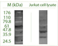
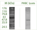
Anti-Autophagy-related protein 16-1 (ATG16 L1) Antibody
Our Anti-Autophagy-related protein 16-1 (ATG16 L1) rabbit polyclonal primary antibody detects human and rat Autophagy-related protein 16-1 (ATG16 L1), and is whole serum. It is validated for use in ELISA, ICC, WB.
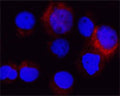
Atg16 L1 detected in cytospun HL60 cells by fluorescence confocal microscopy using antibody R-158-100. HL60 cells were fixed with acetone 90 seconds; primary antibody was diluted 1:100 and incubated for 1 hour, 3 x washes and secondary (Goat anti Rb 1:100 Alexa 546 conjugate) for 30 minutes. Cells stained for Atg16 L1 appear in red. The cells were then counter-stained with Hoechst Dye (blue colour).
Click on image to zoom
SKU: R-158-100
Ships: 1-2 business days
Product Details
Autophagy-related protein 16-1 (ATG16 L1)
ATG16 L1 plays an essential role in autophagy. It is a homooligomer ant interacts with ATG5. Part of either the minor and major complexes respectively composed of 4 sets of ATG12-ATG5 and ATG16L1 (400 kDa) or 8 sets of ATG12-ATG5 and ATG16L1 (800 kDa). APG16 L1 localises to the cytoplasm, in particularly to preautophagosomal structure (PAS) where it is involved in the membrane targeting of ATG5. There are 5 named isoforms produced by alternative splicing. Genetic variation within ATG16 L1 are associated with susceptibility to inflammatory bowel disease type 1.
Whole serum
Polyclonal
Mixed
ELISA, ICC, WB
Rabbit
22 kDa
N-terminal of human ATG16-L1 protein conjugated to Blue Carrier Protein has been used as the immunogen.
Human
Human, Rat
Spin vial briefly before opening. Reconstitute in 100 µL sterile-filtered, ultrapure water. Centrifuge to remove any insoluble material. After reconstitution keep aliquots at -20°C for a higher stability, and at 2-8°C with an appropriate antibacterial agent. Avoid repetitive freeze/thaw cycles.
Lyophilized
WB: 1:1000-1:4000
IHC: 1:200-1:1000
ELISA: 1:1000-1:4000
IHC: 1:200-1:1000
ELISA: 1:1000-1:4000
Unconjugated
Detects human protein via non-reduced WB, and ELISA human, rat. Other species not yet tested.
For research use only.
United States
12 months after date of receipt (unopened vial).
APG16L; Autophagy-related protein 16-1; ATG16L; APG16-like 1; IBD10; ATG16L1; WDR30; APG16L beta; ATG16 autophagy related 16-like 1; WD repeat domain 30
Q676U5
UniProt Summary: Plays an essential role in both canonical and non-canonical autophagy: interacts with ATG12-ATG5 to mediate the lipidation to ATG8 family proteins (MAP1LC3A, MAP1LC3B, MAP1LC3C, GABARAPL1, GABARAPL2 and GABARAP). Acts as a molecular hub, coordinating autophagy pathways via distinct domains that support either canonical or non-canonical signaling. During canonical autophagy, interacts with ATG12-ATG5 to mediate the conjugation of phosphatidylethanolamine (PE) to ATG8 proteins, to produce a membrane-bound activated form of ATG8. Thereby, controls the elongation of the nascent autophagosomal membrane. As part of the ATG8 conjugation system with ATG5 and ATG12, required for recruitment of LRRK2 to stressed lysosomes and induction of LRRK2 kinase activity in response to lysosomal stress. Also involved in non-canonical autophagy, a parallel pathway involving conjugation of ATG8 proteins to single membranes at endolysosomal compartments, probably by catalyzing conjugation of phosphatidylserine (PS) to ATG8. Non-canonical autophagy plays a key role in epithelial cells to limit lethal infection by influenza A (IAV) virus. Regulates mitochondrial antiviral signaling (MAVS)-dependent type I interferon (IFN-I) production. Negatively regulates NOD1- and NOD2-driven inflammatory cytokine response. Instead, promotes an autophagy-dependent antibacterial pathway together with NOD1 or NOD2. Plays a role in regulating morphology and function of Paneth cell.
55054
25°C (ambient)