
Acridine Orange Fluorescent Staining Solution

For fluorescence microscopy and flow cytometry analysis of cellular physiology and cell cycle status. It may be used in conjunction with other stains / fluorogenic substrates including our Magic Red® products. Can also be used as a bacterial stain.
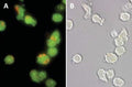
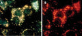
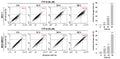
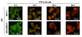

Figure 1. Normal Jurkat cells stained with Acridine Orange (AO) show orange lysosomal staining. Cells were stained with 5 µM AO in PBS for 60 minutes at 37°C. Photomicrographs were taken using a Nikon Eclipse E800 photomicroscope using a 460-500 nm excitation filter and a 505-560 nm emission / barrier filter set at 300X. Photo B shows the corresponding DIC image of the cells in A (AO appears faintly). Data courtesy of Dr. Zbigniew Darzynkiewicz (Brander Cancer Research Institute, New York, NY).
Click on image to zoom



Ships: 1-2 business days
- AO may be used neat or diluted in diH2O, PBS, or media. ICT’s AO is supplied at 0.5 mL liquid at 1 mM (266 µg/mL). 2. Add AO to the cell sample media at 0.5-5 µM, equal to a final dilution of 1:2,000 – 1:200 in the cells (.05-.5% v/v). For example, if using AO at 1.0 µM in the final cell suspension, it must be diluted 1:1,000. First dilute it 1:100 in PBS or diH2O; e.g., put 10 µL AO into 990 µL PBS or diH2O. Pipette the diluted AO into the cell suspension at approximately 1:10; e.g., put 50 µL diluted AO into 450 µL cell suspension.
- Incubate 15-30 minutes at 37°C.
- Wash cells if reagent is too bright.
- Analyze with fluorescence. Lysosomes will appear yellowish green by illuminating cells with a blue light (488 nm) excitation filter and a green light (540-550 nm) emission/barrier filter. Alternatively, lysosomes will appear red when using an excitation filter of 550 nm (540-560 nm) and a long pass >610 nm emission/barrier filter.