Anti-Microtubule Associated Protein 2 (MAP2) Antibody (5H11)

Our Anti-Microtubule Associated Protein 2 (MAP2) mouse monoclonal primary antibody detects bovine, human, mouse, and rat Microtubule Associated Protein 2 (MAP2), and is IgG. It is validated for use in ICC, IHC-Frozen, WB.
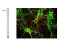
![Left: Rat hippocampus section stained by Immunohistochemistry with mouse anti-MAP2 (green, 1:5,000) and rabbit antibody to alpha-internexin (R-1379-50, red, 1:2,000). IHC Method: Following transcardial perfusion of rat with 4% paraformaldehyde, brain was post-fixed for 24 hours, cut to 45 um, and free-floating sections were stained. MAP2 protein is detected in the perikarya and dendrites of the most neurons, and the alpha-internexin antibody selectively stains axons and dendrites of neuronal cells. Right: Western blot analysis of tissue lysates using mouse anti-MAP2 (green, 1:10,000). [1] protein standard, [2] adult rat whole brain, [2] embryonic (E20) rat brain, [4] adult rat spinal cord, and [5] adult mouse brain. A band at about 280 kDa corresponds to full length MAP2A and MAP2B protein. MAP2A/B is expressed heavily in adult brain particularly in cortical regions, but is a more minor component of spinal cord and almost absent from the embryonic brain sample. Note that the epitope for this antibody in within the projection domain found only in MAP2A and MAP2B, thus the antibody does not bind to the lower molecular weight MAP2C and MAP2D isoforms which lack this region.](http://www.antibodiesinc.com/cdn/shop/files/m-1625-100-ihc-wb_120x120.jpg?v=1759273303)
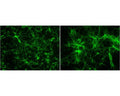

Whole rat brain lysate with mouse anti-MAP2 antibody. The antibody recognizes the ~280 kDa protein. Right: Mixed neuron/glia cultures stained with mouse anti-MAP2 (green) and also rabbit antibody to neurofilament H (Catalog Number R-1388-50) (red). Since the NF-H protein is largely expressed in neuronal axons, while the MAP2 is only found in neuronal dendrites and perikarya, there is little overlap between these two staining patterns. DNA stain shows nuclei of neurons and non-neuronal cells (blue).
Click on image to zoom
Ships: 1-2 business days