Anti-Vimentin Antibody
Our Anti-Vimentin chicken polyclonal primary antibody detects human, mouse, other mammals (predicted), and rat Vimentin, and is IgY preparation. It is validated for use in ICC, IHC-Frozen, WB.
![Left: Analysis of vimentin expression in HeLa cells by Immunocytochemistry. Cells were stained with chicken antibody to vimentin (1:10,000, green), and co-stained with a mouse anti-actin antibody (red). Blue: DAPI nuclear stain. The vimentin antibody stains the intermediate filament network, while the actin antibody labels the submembranous cytoskeleton, stress fibers, and bundles of actin associated with cell adhesion sites. Right: Western blot analysis of tissue and cell lysates using chicken antibody to Vimentin (1:5,000, red, lanes 2-6). [1] protein standard, [2] rat whole brain, [3] HeLa, [4] SH-SY5Y, [5] HEK293, [6] NIH-3T3. Vimentin protein appears as single band at around 50 kDa. The blot was simultaneously probed with a mouse antibody to MAP2C/D (green).](http://www.antibodiesinc.com/cdn/shop/files/c-1409-50-ihc-wb_120x120.jpg?v=1759274040)

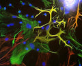
View of mixed neuron/glial cultures stained with Chicken polyclonal antibody to Vimentin C-1409-50 (green) and Rabbit polyclonal antibody to Glial Fibrillary Acidic Protein R-1374-50 (red). Vimentin is expressed alone in fibroblastic and endothelial cells, which are the flattened cells in the middle of the imate which appear green. Astrocytes may express primarily Glial Fibrillary Acidic Protein (GFAP), or GFAP and vimentin, and so appear red (GFAP only) or golden yellow (GFAP and Vimentin). In cells which express both GFAP and vimentin, the two protein assemble to produce heteropolymer filaments.
Click on image to zoom
Ships: 5-7 business days