Anti-Ubiquilin 2 Antibody (6H9)
Our Anti-Ubiquilin 2 mouse monoclonal primary antibody detects human and mouse Ubiquilin 2, and is IgG. It is validated for use in ICC, WB.


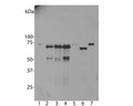
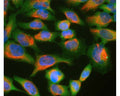
![Left: Analysis of ubiquilin 2 expression in NIH-3T3 cell culture by Immunocytochemistry. Cells were stained with mouse antibody to ubiquilin 2 (green, 1:1,000), and co-stained with chicken antibody to lamin A/C (C-1698-100, red, 1:5,000). Blue: DAPI nuclear stain. Cells were treated with 50 uM of chloroquine, an inhibitor of autophagy, for 16 hours prior to staining. The ubiquilin 2 antibody reveals punctate staining of ubiquilin 2 protein accumulated in lysosomes in the cytoplasm, while the lamin A/C antibody stains the nuclear lamina. Right: Western blot analysis of tissue and cell lysates using mouse antibody to ubiquilin 2 (green, 1:1,000). [1] protein standard, [2] NIH-3T3, [3] C6, [4] HEK293, [5] HeLa, [6] SH-SY5Y, [7] rat whole brain, and [8] mouse whole brain. The band at 65-70 kDa corresponds to ubiquilin 2 protein, which is known to differ between human and rodent species.](http://www.antibodiesinc.com/cdn/shop/files/m-1656-100-ihc-wb_120x120.jpg?v=1759273341)
Western blot analysis of untransfected primary mouse neuron and glia cell cultures (lane 1), the same cells transduced with human ubiquilin 2 wild type (lane 2), with ubiquilin 2 P506T mutant (lane 3), with ubiquilin 2 P497S mutant (lane 4), with enchanced GFP control (lane 5), in HeLa cells (lane 6) and 3T3 cells (lane 7).
Click on image to zoom
SKU: M-1656-100
Product Details
Ubiquilin 2
Ubiquilin 2 (also known as PLIC2 and Chap1) is a member of the ubiquilin protein family, which regulate the degradation of cellular proteins through proteasome or autophage-like pathways (1, 2, 3). Humans have four ubiquilin genes, each encoding a separate protein referred to as Ubiquilin 1, 2, 3 and 4. All ubiquilins contain an N-terminal ubiquitin-like (UBL) domain and a C-terminal ubiquitin-associated (UBA) domain, while the central part of the molecules are highly variable. The UBL domains bind subunits of the proteasome, and the UBA domains binds to polyubiquitin chains that are typically conjugated onto proteins marked for proteosomal degradation (1). Ubiquilin 2 has a unique region close to the C terminus containing 12 PXX tandem collagen like repeats, where P is proline and X is most cases valine, glycine, isoleucine or threonine. Teepu Siddique and his collaborators have identified mutations in the ubiquilin 2 gene leading to protein point mutations which were important contributors to several forms of amyotrophic lateral sclerosis (ALS) and Frontotemporal lobar degeneration (FTLD). Interestingly, these mutations involved alterations in proline residues in the PXX repeat region (P497H, P497S, P506T, P509S and P525S, ref. 4). Recently, the Lee and Trojanowski group investigated C9orf72 hexanucleotide expansion and ubiquilin 2 pathology in patients with ALS and FTLD by genetic analysis and immunohistochemistry and found distinct ubiquilin 2 pathology in ALS and FTLD-TDP with C9orf72 expansion (5). C9orf72 hexonucleotide expansion is the most commmon cause to date of familial ALS and FTLD (6, 7). Ubiquilin 2 protein is of different molecular size in mouse and human, 638 and 624 amino acids respectively. As a result the mouse protein, endogenously expressed in rodent 3T3 cells, runs on SDS-PAGE and western blots slightly slower than the human protein.
IgG
Monoclonal
6H9
IgG1
ICC, WB
Mouse
~66 kDa in human Hela cells and 68 kDa in rodent 3T3 cells
Recombinant human ubiquilin 2 expressed and purified from E. coli.
Human
Human, Mouse
Spin vial briefly before opening. Reconstitute with 100 µL sterile-filtered, ultrapure water to achieve a 1 mg/mL concentration. Centrifuge to remove any insoluble material. Aliquot and store at -20°C for up to six months after date of receipt. Avoid freeze-thaw cycles.
Lyophilized
Protein G purified
Lyophilized from PBS buffer pH 7.2-7.6 with 0.1% trehalose, and sodium azide
WB: 1:1000-1:2000
ICC: 1:500-1:1000
ICC: 1:500-1:1000
Unconjugated
In primary mouse neuron and glia cell culture, endogenous ubiquilin 2 appears as a weak band at 68 kDa in all tranduced and non-transduced cells, indicating low endogenous expression of mouse ubiquilin 2. Strong bands are seen in cells transduced with human wild type or mutant ubiquilin 2. Small proteins which run at 50 kDa in these cells are the fragments of ubiquilin 2. Note, ubiquilin 2 runs at ~66 kDa in human Hela cells and 68 kDa in rodent 3T3 cells. The antibody has also been used successfully for immunocytochemistry.
For research use only.
United States
12 months after date of receipt (unopened vial).
25°C (ambient)