Anti-Rhodopsin Antibody (B630)
Our Anti-Rhodopsin mouse monoclonal primary antibody detects bovine, human, mouse, pig, and rat Rhodopsin, and is affinity-purified. It is validated for use in IHC-Frozen, WB.

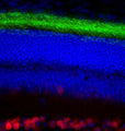
Left: Pig retinal section stained for rhodopsin by Immunohistochemistry with mouse antibody to Rhodopsin (green). Rhodopsin is most abundant in the rod outer segments (ROS) of retina, clearly localized in rod membranes. The rod inner segments (RIS) and rod nuclei in the outer nuclear layer (ONL) are also seen in this image. Nuclear DNA was stained with DAPI (blue). Right: Western Blot of bovine retinal extract probed with mouse antibody to Rhodopsin. The antibody stains a band corresponding to retinal rhodopsin at about 35 kDa. Higher molecular weight bands represent aggregated forms of rhodopsin.
Click on image to zoom
SKU: M-1815-100
Product Details
Rhodopsin
Photoreceptor required for image-forming vision at low light intensity. Required for photoreceptor cell viability after birth (By similarity). Light-induced isomerization of 11-cis to all-trans retinal triggers a conformational change that activates signaling via G-proteins (PubMed:10926528, PubMed:12044163, PubMed:11972040, PubMed:16908857, PubMed:16586416, PubMed:17060607, PubMed:17449675, PubMed:18818650, PubMed:21389983, PubMed:22198838, PubMed:23579341, PubMed:25205354, PubMed:27458239). Subsequent receptor phosphorylation mediates displacement of the bound G-protein alpha subunit by the arrestin SAG and terminates signaling (PubMed:1396673, PubMed:15111114). Ref: uniprot.org
Affinity-purified
Monoclonal
B630
IgG1
IHC, WB
Mouse
Purified bovine rhodopsin
Bovine
Bovine, Human, Mouse, Pig, Rat
Spin vial briefly before opening. Reconstitute with 100 µL sterile-filtered, ultrapure water to achieve a 1 mg/mL concentration. Centrifuge to remove any insoluble material. Store lyophilized antibody at 2-8°C. After reconstitution divide into aliquots and store at -20°C for long-term storage. Store at 2-8°C short-term (up to 4 weeks) with an appropriate antibacterial agent. Avoid repetitive freeze/thaw cycles.
Lyophilized
Protein G purified
Lyophilized from PBS buffer pH 7.2-7.6 with 0.1% trehalose, and sodium azide.
WB: 1:5000
IHC: 1:1000
IHC: 1:1000
Due to the highly hydrophobic nature of rhodopsin, avoid boiling samples in SDS-PAGE sample buffer for rhodopsin analysis by Western Blotting, as this will result in extensive aggregation of the rhodopsin protein and appearance of high molecular weight bands.
Unconjugated
Bovine, reacts with human, rat, mouse, cow, pig. Expected to react with other mammalian species.
For research use only.
United States
12 months after date of receipt (unopened vial).
25°C (ambient)