Anti-NGFR/p75 neurotrophin receptor (p75NTR) Antibody (8J2)
Our Anti-NGFR/p75 neurotrophin receptor (p75NTR) mouse monoclonal primary antibody detects human, mouse, and rat NGFR/p75 neurotrophin receptor (p75NTR), and is IgG. It is validated for use in FC, ICC, IHC-Frozen, Immunopanning, IP, WB.
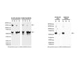
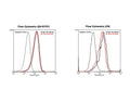
Left: Analysis of p75NTR expression in human and rodent RIPA cell lysates by Western Blotting. Mouse antibody to p75NTR clone 8J2 (M-1818-100) and clone MLR2 (M-009-100) detect p75NTR-IR at ~50-60 kDa in mouse p75NTR-transfected cells (A), but not in non-transfected control cells (B). p75NTR-IR is also observed in human SH-SY5Y (C) and rat C6 (D) cell lysates. Dependent on cell lysate, dimers or trimers (~150 kDa) of p75NTR are detected (Anastasia et al., 2015). 50 ug of protein were loaded per lane. WB Method: SDS-PAGE: 4-12%, non-reducing conditions; Transfer: Tris-Glycine buffer; Membrane: nitrocellulose (0.45 um); Blocking: 5% skim milk in TBST, 1 hour at RT; Primary antibody: 1 µg/mL, overnight at 4°C; Secondary antibody: anti-mouse-HRP (1/6000) 1 hour at RT; Detection: Chemiluminiscence. Right: Immunoprecipitation of p75NTR from rat C6 cell lysate and detection by Western Blotting. Mouse antibody to p75NTR clone 8J2 (M-1818-100) and clone MLR2 (M-009-100) precipitate bands at the expected molecular weight of ~50-60 kDa for p75NTR (A). No p75NTR-IR is observed in remaining supernatant (B) or control (Protein G only, C). IP Method: C6 lysates were prepared in non-denaturing lysis buffer and pre-cleared with Protein G agarose beads (40 uL bead slurry per 1 mg total protein). Cleared lysate was then incubated with either M-1818-100 or M-009-100 (10 ug antibody per 200 ug total protein), and immune complexes bound by Protein G agarose (10 uL). Precipitated p75NTR was eluted off the beads and antibody by heating and addition of SDS-PAGE sample buffer. WB Method: as above, with primary goat anti-p75NTR (1 µg/mL) antibody and secondary anti-sheep-HRP (1/6000) antibody. Detection: Chemiluminiscence.
Click on image to zoom
SKU: M-1818-100
Product Details
NGFR/p75 neurotrophin receptor (p75NTR)
p75NTR (CD271) was originally discovered as a low affinity nerve growth factor receptor (NGFR). Later it was found that it was the receptor for all neurotrophins, including NGF, BDNF, NT3 and NT4/5. It mediates signals of neurotrophins for neuronal survival, apoptosis, neurite outgrowth and synaptic plasticity. Recently, it has been revealed that p75NTR not only acts as the receptor for neurotrophins but also the receptor for many other pathological ligands such as prions, rabies virus and amyloid beta. p75NTR also acts as a co-receptor for NOGO which mediates inhibitory signals of myelin associated protein. p75NTR is highly expressed in a number of non-neuronal and neuronal cells including motor neurons during development and also in damaged neurons. Recent research proposes the extracellular domain of p75NTR as a biomarker for monitoring the progression of motor neuron disease (MND), also known as Amyotrophic Lateral Sclerosis (ALS) or Lou Gehrig's Disease. SUBUNIT: Homodimer; disulfide-linked. Interacts with p75NTR-associated cell death executor. Interacts with NGFRAP1/BEX3.
IgG
Monoclonal
8J2
IgG2a
Flow, ICC, IHC, IP, WB
Mouse
Recombinant extracellular domain (amino acids 29-250) of human NGFR/p75NTR protein with N-terminal His-tag.
Human
Human, Mouse, Rat
Spin vial briefly before opening. Reconstitute in 100 µL sterile-filtered, ultrapure water to achieve a concentration of 1 mg/mL. Centrifuge to remove any insoluble material. Final buffer contains no preservatives. Store lyophilized antibody at 2-8°C. After reconstitution divide into aliquots and store at -20°C for long-term storage. Store at 2-8°C short-term (up to 4 weeks) with an appropriate antibacterial agent. Avoid repetitive freeze/thaw cycles.
Lyophilized
Protein A purified IgG
Lyophilized from a solution containing PBS buffer pH 7.2-7.6 with 0.1% trehalose, without preservatives.
WB: 0.5-2.0 µg/mL, non-reducing conditions only (no DTT or beta-mercaptoethanol).
ICC: 1-5 µg/mL
IP: Lysate dependent. 10 ug per 200-500 ug total protein
ICC: 1-5 µg/mL
IP: Lysate dependent. 10 ug per 200-500 ug total protein
Flow Cytometry: 5-20 µg/mL.
Immunopanning: 1-5 µg/mL.
ICC: Staining is strongest in non-fixed cells, light fixation is tolerable.
IHC: fresh, acetone fixed sections only, epitope is fixation sensitive. Not suitable in formalin-fixed, paraffin (FFPE) embedded tissues.
Immunopanning: 1-5 µg/mL.
ICC: Staining is strongest in non-fixed cells, light fixation is tolerable.
IHC: fresh, acetone fixed sections only, epitope is fixation sensitive. Not suitable in formalin-fixed, paraffin (FFPE) embedded tissues.
Unconjugated
Reacts with human, mouse and rat. Cross-reactivity with other species not tested but expected.This antibody is specific for NGFR/p75NTR as demonstrated by Western blotting and immunprecipitation. The antibody recognizes extracellular p75NTR under non-reducing conditions.
For research use only.
United States
12 months after date of receipt (unopened vial).
Low-affinity nerve growth factor receptor; NGF receptor; Gp80-LNGFR; p75 ICD; Low affinity neurotrophin receptor p75NTR; CD271
25°C (ambient)

