Anti-Neurofilament heavy polypeptide, phosphorylated, (pNF-H) Antibody
Our Anti-Neurofilament heavy polypeptide, phosphorylated (pNF-H) chicken polyclonal primary antibody detects bovine, dog, horse, human, mouse, other mammals (predicted), pig, and rat Neurofilament heavy polypeptide, phosphorylated (pNF-H), and is IgY preparation. It is validated for use in ELISA, IF, ICC, IHC, WB.
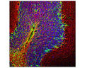
Image of a rat cerebellum section by Immunofluorescence and Immunohistochemistry. The section was stained with C-1386-50, Neurofilament heavy polypeptide, phosphorylated (pNF-H), Chicken pAb, (red, 1:5,000) and co-stained with product R-1374-50, Glial Fibrillary Acidic Protein (GFAP), Rabbit pAb (green). The blue is DAPI staining of nuclear DNA. Method: following transcardial perfusion with 4% paraformaldehyde, brain was post fixed for 24 hours, cut to 45 uM, and free floating sections were stained. The NF-H antibody labels network of axons of different neurons, while the GFAP antibody stains astrocytes.
Click on image to zoom
Bovine, Canine, Horse, Human, Mouse, Pig, Rat
ELISA, ICC, IF, IHC, WB
Chicken
SKU: C-1386-50
Product Details
Neurofilament heavy polypeptide, phosphorylated (pNF-H)
Neurofilaments are the 10 nm or intermediate filament proteins found specifically in neurons, and are composed predominantly of three major proteins called NF-L, NF-M and NF-H, though other filament proteins may be included also. The major function of neurofilaments is likely to control the diameter of large axons. NF-L is the neurofilament light or low molecular weight polypeptide and runs on SDS-PAGE gels at 68-70 kDa with some variability across species. Antibodies to NF-L are useful for identifying neuronal cells and their processes in cell culture and sectioned material. NF-L antibody can also be useful for the visualization of neurofilament rich accumulations seen in many neurological diseases, such as Lou Gehrig's disease (ALS), giant axon neuropathy, Charcot-Marie Tooth disease and others. (Ref: uniprot.org)
IgY preparation
Polyclonal
IgY
ELISA, ICC, IF, IHC, WB
Chicken
200 kDa
This antibody has been made against biochemically isolated NF-H purified from bovine spinal cord.
Bovine
Bovine, Canine, Horse, Human, Mouse, Pig, Rat
Spin vial briefly before opening. Reconstitute with 50 µL sterile-filtered, ultrapure water. Centrifuge to remove any insoluble material. Store lyophilized antibody at 2-8°C After reconstitution of lyophilized antibody, aliquot and store at -20°C for a higher stability. Avoid freeze-thaw cycles. Store at 4°C for up to one month for short term storage and frequent use.
Lyophilized
Lyophilized IgY preparation, with sodium azide.
WB: 1:20000-1:50000
IHC: 1:20000
ICC: 1:20000
IHC: 1:20000
ICC: 1:20000
Unconjugated
Species cross-reactivity includes human, rat, mouse, cow, pig, horse and dog. This antibody reacts with phosphorylated NF-H and is seen as a band at approx 200 kDa in WB. Refer to publication by Shaw et al (2005) for the use of this antibody in an ELISA to detect NF-H. Predicted to react with other mammalian tissues due to sequence homology.
For research use only.
United States
12 months after date of receipt (unopened vial).
NF-H; NFH; NF-200; NF200; NF-H; NEFH; N52; Neurofilament heavy polypeptide; Neurofilament triplet H protein; 200 kDa neurofilament protein; KIAA0845
25°C (ambient)

