Anti-Microtubule-associated protein tau (MAPT) Antibody (2E9)
Our Anti-Microtubule-associated protein tau (MAPT) mouse monoclonal primary antibody detects human, mouse, other mammals (predicted), and rat Microtubule-associated protein tau (MAPT), and is affinity-purified. It is validated for use in ICC, WB.
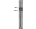
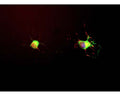
![Left: Analysis of MAP-tau expression (green) in cortical neuron-glial culture from E20 rat by Immunocytochemistry. Mouse antibody to MAP-tau was used at 1:1,000 dilution. Co-staining was performed with chicken antibody to MAP2 (C-1382-50, red, 1:5,000). Blue: DAPI nuclear stain. The MAP-tau antibody stains perikarya, dendrites and axons of neurons, while the MAP2 antibody labels only dendrites and perikarya. As a result, perikarya and dendrites appear orange-yellow, since they contain both proteins. Right: Western blot analysis of MAP-tau expression in tissue lysates using mouse antibody to MAP-tau (green, 1:2,000). [1] protein standard, [2] rat brain, [3] rat spinal cord, [4] mouse brain, [5] mouse spinal cord. Tau protein is expressed as up to 9 different isoforms of different molecular weights, therefore, it appears as multiple closely spaced bands in the range from 48 kDa to 67 kDa in the CNS (lanes 2-4). In the PNS, additional higher molecular weight bands are observed (lane 5).](http://www.antibodiesinc.com/cdn/shop/files/m-1703-100-ihc-wb_120x120.jpg?v=1759273423)

Crude rat brain extract. Tau protein is expressed as up to 9 different isoforms of different molecular weight and so appears as multiple closely spaced bands covering the region of the blot from 48 kDa to 67 kDa.
Click on image to zoom
Ships: 1-2 business days