Anti-Lysosomal Associated Membrane Protein 1 (LAMP1) Antibody (5H6)
Our Anti-Lysosomal Associated Membrane Protein 1 (LAMP1) mouse monoclonal primary antibody detects human Lysosomal Associated Membrane Protein 1 (LAMP1), and is IgG. It is validated for use in FC, ICC, WB.
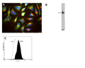
![Left: Detection of LAMP1 protein in HeLa cells by Immunocytochemistry. LAMP1 was stained with mouse antibody to LAMP1 (red, 1:500), and co-stained with chicken antibody to vimentin (C-1409-50, green, 1:10,000). Blue: DAPI nuclear stain. The cells were treated with 50 uM chloroquine, an inhibitor of autophagy, for 16 hours prior to staining. The LAMP1 antibody reveals vesicular staining of LAMP1 protein accumulated in swollen lysosomes, while the vimentin antibody specifically labels the intermediate filament network in these cells. Right: Western blot analysis of LAMP1 protein expression in cell lysates using mouse antibody to LAMP1 (green, 1:10,000). Cells were maintained under normal conditions (Ct), or treated with 50 uM chloroquine (CQ), an inhibitor of autophagy, for 24 hours: [1] protein standard, [2] HeLa Ct, [3] HeLa+CQ, [4] NIH-3T3 Ct, and [5] NIH-3T3+CQ. The smeared band between 75-120 kDa rresponds to variably glycosylated forms of the LAMP1 protein detected only in the human cells, this antibody does not recognize the rodent LAMP1 homologue. The same blot was probed with a chicken antibody to HSP60 (red, lanes 2-5).](http://www.antibodiesinc.com/cdn/shop/files/m-1690-100-ihc-wb_120x120.jpg?v=1759273393)

A: HeLa cell staining with M-1690-100 (red), and counterstained with chicken polyclonal antibody to Vimentin C-1409-50 (green) and DNA (blue). The LAMP1 antibody reveals strong punctate cytoplasmic staining corresponding to lysosomes and late endosomes, while the Vimentin antibody reveals cytoplasmic intermediate filaments. B: Western blot of HeLa cell crude extract stained with M-1690-100 (lane 9). The antibody binds to a diffuse band running at between 90 and 120 kDa. C: Analysis of LAMP1 expression in human neuroblastoma SH-SY5Y cell line by Flow Cytometry. Fixing and Permeabilization of cells: Absolute methanol (10 minutes in ice) and 0.1% Tween-20 in PBS, Blocking: 1% BSA, Primary antibody: Mouse Monoclonal antibody to LAMP1 (cat # M-1690-100, 2μg per ~10^6 cells) for 30 minutes at room temperature, Secondary antibody: Goat anti-mouse PE labeled secondary antibody (1:100 fold dilution) with incubation for 20 minutes in dark at room temperature. Non-specific Control IgG, clone X63 (cat # M-1249-100) was used as negative control under same conditions (black dashed). Flow cytometry data and results were generated using Orflo MoxiflowTM instrument and protocols.
Click on image to zoom
Ships: 5-7 business days