Anti-Lamin A/C Antibody (4C4)
Our Anti-Lamin A/C mouse monoclonal primary antibody detects human Lamin A/C, and is IgG. It is validated for use in FC, ICC, WB.
![Left: Lamin A/C expression in HeLa cells, stained with mouse antibody to lamin A/C (red, 1:2,000) by Immunocytochemistry. Co-staining was performed with a rabbit anti-HSP60 antibody (green). Blue: Hoechst nuclear stain. The lamin A/C antibody specifically labels the nuclear lamina. Right: Western blot analysis of lamin A/C expression in cell lysates using mouse anti-lamin A/C antibody (green, 1:1,000). [1] protein standard, [2] HeLa, [3] HEK293 [4] C6, and [5] NIH-3T3 cell lysates. Two strong bands at 74 and 65 kDa correspond to the lamin A and lamin C proteins, respectively, detected only in the cells of human origin. The lamin A/C antibody does not recognize rat or mouse proteins.](http://www.antibodiesinc.com/cdn/shop/files/m-1689-100-ihc-wb_120x120.jpg?v=1759273386)

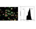
Left: HeLa cells stained with M-1689-100 (red), and counterstained with chicken polyclonal antibody to Vimentin (green, C-1409-50). The The Lamin A/C antibody reveals strong nuclear lamina staining, while the Vimentin antibody reveals cytoplasmic intermediate filaments. The blue stain reveals DNA in the nuclei of these cells. Right: Analysis of Lamin A/C expression in human prostate cancer DU145 cell line by FLow Cytometry. Fixing and Permeabilization of cells: Absolute methanol (10 minutes in ice) and 0.1% Tween-20 in PBS, Blocking: 200 µg/mL Normal Sheep IgG (20 minutes), Primary antibody: Mouse Monoclonal antibody to Lamin A/C (cat # M-1689-100, 2 μg per ~10^6 cells) for 30 minutes at room temperature, Secondary antibody: Goat anti-mouse PE labeled secondary antibody (1:100 fold dilution) with incubation for 20 minutes in dark at room temperature. Non-specific Control IgG, clone X63 (cat # M-1249-100) was used as negative control under same conditions (black dashed). Flow cytometry data and results were generated using Orflo MoxiflowTM instrument and protocols.
Click on image to zoom
Ships: 1-2 business days