Anti-Coronin-1A Antibody
Our Anti-Coronin-1A rabbit polyclonal primary antibody detects bovine, human, mouse, pig, and rat Coronin- 1A, and is whole serum. It is validated for use in ICC, WB.
![Left: Detection of Coronin 1a (red) in cortical neuron-glial cell culture from E20 rat by Immunocytochemistry. Coronin 1a antibody was used at 1:1,000 dilution. Cells were co-stained with mouse antibody to GFAP (M-1375-100, green, 1:1,000). Blue: Hoechst nuclear stain. The coronin 1a antibody labels protein expressed in the cytoplasm of microglia cells, while the GFAP antibody stains intermediate filaments in astrocytic cells. Right: Western blot analysis of coronin 1a expression in tissue lysates using rabbit anti-coronin 1a antibody (green, 1:5,000). [1] protein standard, [2] mouse brain, [3] rat brain, [4] cow cerebellum, [5] cow cortex, and [6] pig spinal cord. The strong single band about 55 kDa corresponds to the coronin 1a protein.](http://www.antibodiesinc.com/cdn/shop/files/r-1335-50-ihc-wb_120x120.jpg?v=1759273896)

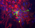
Immunocytochemistry of a mixed neuron/glial culture from newborn rat brain stained with Rabbit polyclonal antibody to Coronin 1a R-1335-50 (green) at a dilution of 1:10,000 and Chicken polyclonal antibody to Vimentin C-1409-50 at 1:5,000. Blue is nuclear DNA counter stain. Glial cells and fibroblasts stain with Vimentin, while microglia alone stain strongly and specifically for Coronin 1a, which can therefore be used as a robust marker of this important cell type.
Click on image to zoom
Ships: 1-2 business days