Anti-Contactin-6 Antibody
Our Anti-Contactin-6 rabbit polyclonal primary antibody detects human, mouse, and rat Contactin-6, and is whole serum. It is validated for use in IHC-Frozen.
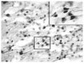
Immunohistochemical detection of contactin-6 in rat sciatic nerve using antisera R-023-50. Anesthetized animals were perfused with 4% paraformaldehyde in PBS containing 12.5% picric acid. Floating cryostat sections of sciatic nerve were permeabilized with 0.5% NP-40 in Tris-buffered saline and blocked with 5% normal goat serum. Sections were incubated with polyclonal rabbit antibody to contactin-6 diluted 1:3000, followed by incubation with biotinylated sheep anti-rabbit antibody and avidin-biotin-peroxidase complex. The immunocomplex was visualized with DAB.
Click on image to zoom
SKU: R-106-100
Product Details
Contactin-6
The neural adhesion molecule Contactin-6, also known as NB-3, is a contactin/F3 subgroup member of immunoglobulin superfamily. It is expressed exclusively in the nervous system and mainly upregulated at the early postnatal stage during mouse brain development. Employing Northern blot analysis Kamei et al found that amongst different regions of the adult human nervous system cerebellum expressed highest level of NB-3 mRNA. The expression of NB-3 in the cerebellum increases until adulthood. In contrast, the expression in the cerebrum declines to a low level after postnatal day 7. NB-3 like other neural recognition molecules plays a vitally important role in axonal guidance during development, plasticity, and maintenance of synaptic connections in the adult brain. Cui et al recently showed that NB-3 acts as a novel Notch ligand to participate in oligodendrocyte generation. Furthermore, NB-3 triggers nuclear translocation of the Notch intracellular domain and promotes oligodendrogliogenesis from progenitor cells and differentiation of oligodendrocyte precursor cells via Deltex1. In primary oligodendrocytes, NB-3 increases myelin-associated glycoprotein transcripts. Hence, the NB-3/Notch signaling pathway may be worthwhile a closer examination for its potential for the treatment of demyelinating diseases. Human NB-3 shares with rat NB-3 86% identity in nucleotide sequences and 90% identity in amino acid sequences. FUNCTION: Contactins mediate cell surface interactions during nervous system development. Participates in oligodendrocytes generation by acting as a ligand of NOTCH1. Its association with NOTCH1 promotes NOTCH1 activation through the released notch intracellular domain (NICD) and subsequent translocation to the nucleus. Involved in motor coordination. SUBCELLULAR LOCATION: Cell membrane; lipid-anchor; GPI-anchor. ALTERNATIVE PRODUCTS: 2 named isoforms produced by alternative splicing. TISSUE SPECIFICITY: Expressed in brain. In brain, it is preferentially expressed in the accessory olfactory bulb, layers II/III and V of the cerebral cortex, piriform cortex, anterior thalamic nuclei, locus coeruleus of the pons and mesencephalic trigeminal nucleus and in Purkinje cells of the cerebellum.
Whole serum
Polyclonal
Mixed
IHC
Rabbit
A synthetic peptide (GYPSPHYRWKQ) as part of mouse contactin-6 conjugated to KLH has been used as the immunogen.
Mouse
Human, Mouse, Rat
Spin vial briefly before opening. Reconstitute in 100 µL sterile-filtered, ultrapure water. Centrifuge to remove any insoluble material. After reconstitution keep aliquots at -20°C for increased stability, and at 2-8°C with an appropriate antibacterial agent. Avoid repetitive freeze/thaw cycles. Glycerol (1:1) may be added for an additional stability.
Lyophilized
IHC: 1:1000-1:4000
Unconjugated
This antiserum stains beautifully the NB3 in DRG, where it nicely stains a subpopulation of neurons and in the trigeminal nerve root, where staining is restricted to the oligodendrocytes. Confirmed to react with rat Contactin-6 and expected to recognize human and mouse Contactin-6 as well.
For research use only.
United States
12 months after date of receipt (unopened vial).
Neural recognition molecule NB-3; mNB-3
25°C (ambient)

