Anti-Cellular oncogene fos (c-Fos) Antibody (2H2)
Our Anti-Cellular oncogene fos (c-Fos) mouse monoclonal primary antibody detects bovine, chicken, horse, human, mouse, pig, and rat Cellular oncogene fos (c-Fos), and is affinity-purified. It is validated for use in ICC, IHC-Frozen.
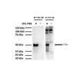
![Left: Rat hippocampus stained for c-FOS (red) by Immunohistochemistry. Section was co-stained with rabbit antibody to FOX3/NeuN (R-3770-100, green). Blue: DAPI nuclear stain. The hippocampal neurons stain green for FOX3/NeuN and a few also are expressing c-FOS, and thus appear orange. These cells were spontaneously active at the time the animal was sacrificed. Right: Western blot analysis of c-FOS expression (green) in cell lysates. Mouse antibody to c-FOS was used at 1:1,000 dilution. GAPDH (red, lanes 2-5) was used as loading control (rabbit antibody to GAPDH, R-1701-100, 1:20,000). [1] protein standard, [2] HeLa cells in serum free media, [3] HeLa cells stimulated with 20% FBS for 2 hours after 36 hours serum starvation, [4] rat cortical neurons, [5] rat cortical neurons treated with membrane depolarization buffer for 5 hours. Multiple bands at 50-65 kDa in stimulated or treated cell lysates correspond to different forms of the c-Fos protein. The single band at 37 kDa (red) represents GAPDH protein.](http://www.antibodiesinc.com/cdn/shop/files/m-1752-100-wb-ihc-2_120x120.jpg?v=1759273489)

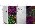
A: Western blot analysis of c-Fos expression in HeLa cells using M-1752-100. HeLa cells were serum-starved for 36 hours (Lane 1). Serum-starved HeLa cells were stimulated with 20% FBS for 2 hours (Lane 2). M-1752-100 recognizes bands in the range of 50-65 kDa, which represent multiple forms of c-Fos. A loading control was performed by stripping and re-probing the membrane with a monoclonal antibody against GAPDH, M-1376-250.
B: Immunofluorescence staining of HeLa cells with M-1752-100. c-Fos staining (green) only localizes in the nuclei of 20% FBS stimulated cells (bottom panel), but not in un-stimulated cells (top panel). Cells were counter-stained with Chicken polyclonal antibody against vimentin, C-1409-50 (red) and DAPI (blue).
C: Immunohistochemistry using M-1752-100 on 4% PFA transcardial-perfused mouse brain sections (45 uM thickness). c-FOS immunoreactive cells (dark colour, localized in cell nucleus) were visualized using a standard HRP-DAB (horseradish peroxidase-3,3'-diaminobenzidine) staining technique.
D: Immunohistochemistry using M-1752-100 (red) and Rabbit polyclonal anti-NeuN/Fox3 (R-3770-100, green) on mouse cortical sections. Neurons positive for c-Fos and Fox3/NeuN appear yellow. The insert shows an enlarged image of staining with M-1752-100. Nuclei were labeled with DAPI (blue).
Click on image to zoom
B: Immunofluorescence staining of HeLa cells with M-1752-100. c-Fos staining (green) only localizes in the nuclei of 20% FBS stimulated cells (bottom panel), but not in un-stimulated cells (top panel). Cells were counter-stained with Chicken polyclonal antibody against vimentin, C-1409-50 (red) and DAPI (blue).
C: Immunohistochemistry using M-1752-100 on 4% PFA transcardial-perfused mouse brain sections (45 uM thickness). c-FOS immunoreactive cells (dark colour, localized in cell nucleus) were visualized using a standard HRP-DAB (horseradish peroxidase-3,3'-diaminobenzidine) staining technique.
D: Immunohistochemistry using M-1752-100 (red) and Rabbit polyclonal anti-NeuN/Fox3 (R-3770-100, green) on mouse cortical sections. Neurons positive for c-Fos and Fox3/NeuN appear yellow. The insert shows an enlarged image of staining with M-1752-100. Nuclei were labeled with DAPI (blue).
Ships: 1-2 business days