Anti-FAM129B (Ser679, 683) Antibody
Our Anti-FAM129B (Ser679, 683) rabbit polyclonal phosphospecific primary antibody from PhosphoSolutions is produced in-house. It detects human and mouse FAM129B (Ser679, 683) and is antigen affinity purified from pooled serum. It is great for use in WB.

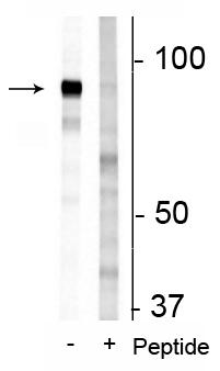
Western blot of 3T3 cell lysate showing specific immunolabeling of the ~83 kDa FAM129B protein phosphorylated at Ser679/683 in the first lane (-). Phosphospecificity is shown in the second lane (+) where immunolabeling is blocked by preadsorption of the phosphopeptide used as the antigen, but not by the corresponding non-phosphopeptide (not shown).
Click on image to zoom
SKU: p170-679
Ships: 1-2 business days
Product Details
FAM129B (Ser679, 683)
FAM129B, also known as Niban-like protein 1, belongs to a poorly characterized protein family with unknown category and function. Increased expression of the Niban gene has been observed in renal carcinomas (Adachi et al., 2004; Sun et al., 2007). Suppression of FAM129B expression in HeLa cells has been seen to promote apoptosis, suggesting that it can modulate cell death signaling, and may be involved in the ER stress response (Sun et al., 2007). FAM129B is also up-regulated in various types of thyroid tumors and Hashimoto’s thyroiditis (Matsumoto et al., 2006). It has been suggested that the MAP kinase dependent phosphorylation of FAM129B is important in controlling melanoma cells, as inhibition of B/Raf/MKK/ERK in melanoma cells represses invasion (Old et al., 2009). It is believed that phosphorylated FAM129B not only derepresses invasion, but also regulates events that promote invasion (Old et al., 2009).
Antigen Affinity Purified from Pooled Serum
Polyclonal
IgG
WB
Rabbit
FAM129B
83 kDa
Synthetic phospho-peptide corresponding to amino acid residues surrounding Ser679/683 of human FAM129B, conjugated to keyhole limpet hemocyanin (KLH).
Human
Human, Mouse
Non-Human Primate
AB_2492091
Storage at -20°C is recommended, as aliquots may be taken without freeze/thawing due to presence of 50% glycerol. Stable for at least 1 year at -20°C.
Liquid
Prepared from pooled rabbit serum by affinity purification via sequential chromatography on phospho and non-phosphopeptide affinity columns.
10 mM HEPES (pH 7.5), 150 mM NaCl, 100 µg per ml BSA and 50% glycerol.
WB: 1:1000
Unconjugated
Specific for endogenous levels of the ~83 kDa FAM129B phosphorylated at Ser679/683. Immunolabeling is blocked by preadsorption with the phosphopeptide used as antigen, but not by the corresponding non-phosphopeptide.
Phosphorylated
Ser679, Ser683
Western blots performed on each lot.
For research use only. Not intended for therapeutic or diagnostic use. Use of all products is subject to our terms and conditions, which can be viewed on our website.
After date of receipt, stable for at least 1 year at -20°C.
bA356B19.6 antibody, C9orf88 antibody, chromosome 9 open reading frame 88 antibody, DKFZP434H0820 antibody, FAM129B antibody, family with sequence similarity 129 member B antibody, FLJ13518 antibody, FLJ22151 antibody, FLJ22298 antibody, hypothetical protein LOC64855 antibody, Meg 3 antibody, Meg-3 antibody, Meg3 antibody, Niban like protein 1 antibody, Niban-like protein 1 antibody, NIBL1_HUMAN antibody, OC58 antibody, OTTHUMP00000022187 antibody, OTTHUMP00000022188 antibody, Protein FAM129B antibody
64855
Blue Ice
- Western Blot Protocol: Download
General References
- Adachi, H., Majima, S., Kon, S., Kobayashi, T., Kajino, K., Mitani, H., Hirayama, Y., Shiina, H., Igawa, M. and Hino, O., 2004. Niban gene is commonly expressed in the renal tumors: a new candidate marker for renal carcinogenesis. Oncogene, 23(19), p.3495. PMID: 14990989
- Sun, G.D., Kobayashi, T., Abe, M., Tada, N., Adachi, H., Shiota, A., Totsuka, Y. and Hino, O., 2007. The endoplasmic reticulum stress-inducible protein Niban regulates eIF2α and S6K1/4E-BP1 phosphorylation. Biochemical and Biophysical Research Communications, 360(1), pp.181-187. PMID: 17588536
- Matsumoto, F., Fujii, H., Abe, M., Kajino, K., Kobayashi, T., Matsumoto, T., Ikeda, K. and Hino, O., 2006. A novel tumor marker, Niban, is expressed in subsets of thyroid tumors and Hashimoto's thyroiditis. Human Pathology, 37(12), pp.1592-1600. PMID: 16949643
- Old, W.M., Shabb, J.B., Houel, S., Wang, H., Couts, K.L., Yen, C.Y., Litman, E.S., Croy, C.H., Meyer-Arendt, K., Miranda, J.G. and Brown, R.A., 2009. Functional proteomics identifies targets of phosphorylation by B-Raf signaling in melanoma. Molecular Cell, 34(1), pp.115-131. PMID: 19362540


