Anti-Cellular oncogene fos (c-FOS) Antibody
Our Anti-Cellular oncogene fos (c-FOS) rabbit polyclonal primary antibody detects human, mouse, and rat Cellular oncogene fos (c-FOS), and is affinity-purified. It is validated for use in ICC, IHC-Frozen, WB.


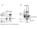
![Western blot analysis of cell lysates using rabbit antibody to c-FOS (green, 1:5,000), and mouse antibody to GAPDH (M-1376-250, red, 1:5,000, lanes 2-5) used as a loading control. [1] protein standard, [2] HeLa cells grown in FBS free media, [3] HeLa cells stimulated with 20% FBS for 2 hours after being in FBS free media for 36 hours, [4] rat cortical neurons, [5] rat cortical neurons treated with membrane depolarization buffer for 5 hours. Multiple bands at 50-65 kDa in stimulated or treated cell lysates correspond to different isoforms of the c-FOS protein.](http://www.antibodiesinc.com/cdn/shop/files/r-1751-50-wb-2_120x120.jpg?v=1759274816)
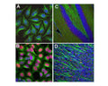
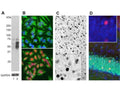
Analysis of c-Fos expression in cultured HeLa cells by Immunocytochemistry (A-B), and in mouse hippocampus (C) or olfactory bulb sections (D) by Immunohistochemistry. HeLa cells were kept in serum-free medium for 36 hours, before cells were stimulated with 20% FBS for 30 minutes (B), while control cells (A) were treated with PBS as a control. The rabbit anti-c-Fos antibody (red, 1:5,000) labels nuclei of stimulated cells, while DAPI (blue) stains all cell nuclei independent of their activity stage. Green: mouse anti-tubulin antibody. Mouse hippocampus (C) and olfactory bulb sections (D) were stained with rabbit anti-c-Fos antibody (red, 1:20,000) and mouse antibody to NF-L (green). The c-FOS antibody stains only nuclei of spontaneously active neurons. NF-L is expressed in axons of neuronal cells. Blue: Cell nuclei stained with DAPI. IHC Method: Following transcardial perfusion of mouse with 4% paraformaldehyde, brain was post fixed for 24 hours, cut to 45 uM, and free-floating sections were stained with above antibodies.
Click on image to zoom
SKU: R-1751-50
Product Details
Cellular oncogene fos (c-FOS)
FUNCTION: Nuclear phosphoprotein which forms a tight but non-covalently linked complex with the JUN/AP-1 transcription factor. In the heterodimer, FOS and JUN/AP-1 basic regions each seems to interact with symmetrical DNA half sites. On TGF-beta activation, forms a multimeric SMAD3/SMAD4/JUN/FOS complex at the AP1/SMAD-binding site to regulate TGF-beta-mediated signaling. Has a critical function in regulating the development of cells destined to form and maintain the skeleton. It is thought to have an important role in signal transduction, cell proliferation and differentiation. In growing cells, activates phospholipid synthesis, possibly by activating CDS1 and PI4K2A. This activity requires Tyr-dephosphorylation and association with the endoplasmic reticulum. SUBUNIT: Heterodimer. Interacts with DSIPI; this interaction inhibits the binding of active AP1 to its target DNA. Interacts with MAFB. SUBCELLULAR LOCATION: Nucleus. INDUCTION: C-fos expression increases upon a variety of stimuli, including growth factors, cytokines, neurotransmitters, polypeptide hormones, stress and cell injury. SIMILARITY: Belongs to the bZIP family. Fos subfamily. SIMILARITY: Contains 1 bZIP domain (Ref: uniprot.org).
Affinity-purified
Polyclonal
IgG
ICC, IHC, WB
Rabbit
50-65 kDa (See application details.)
Full length, E.coli-derived recombinant human c-FOS protein.
Human
Human, Mouse, Rat
Spin vial briefly before opening. Reconstitute with 50 µL sterile-filtered, ultrapure water. Centrifuge to remove any insoluble material. Store lyophilized, unopened vial at 2-8°C or lower. After reconstitution, prepare aliquots and store at -20°C for a higher stability. Avoid freeze-thaw cycles.
Lyophilized
Lyophilized from PBS buffer pH 7.2-7.6 with 0.1% trehalose, and sodium azide
WB: 1:1000-1:2000 (See application details.)
IHC: 1:20000-1:50000 (See application details.)
ICC: 1:2000-1:10000
IHC: 1:20000-1:50000 (See application details.)
ICC: 1:2000-1:10000
IHC: This antibody has been shown to work on 4% PFA fixed mouse brain sections. Note that non-specific staining has been observed on tissue sections when using this antibody at dilutions of 1:5000 or lower. Click for instructions on use of this antibody in IHC on free-floating brain sections.
WB: This antibody detects bands between 50-65 kDa, which only appear in stimulated cells.
Unconjugated
For research use only.
United States
12 months after date of receipt (unopened vial).
Cellular oncogene fos; G0/G1 switch regulatory protein 7; cFOS
25°C (ambient)