Anti-Adenylate cyclase type III (ACIII) Antibody
Our Anti-Adenylate cyclase type III (ACIII) rabbit polyclonal primary antibody detects human (predicted), mouse, other mammals (predicted), and rat Adenylate cyclase type III (ACIII), and is affinity-purified. It is validated for use in ICC, IHC, WB.
![Left: Analysis of rat hippocampus with rabbit antibody to adenylate cyclase III (red) by Immunocytochemistry. Section was co-stained for MeCP2 (green). Blue: DAPI nuclear stain. The ACIII antibody reveals neuronal cilia, while the MeCP2 antibody reveals the neuronal nuclei. Right: Western blot analysis of HEK293 cell lysates using rabbit antibody to ACIII (green, 1:2,000). [1] protein standard, [2] non-transfected HEK293 cells, and [3] HEK293 cells transfected with an expression construct containing a Myc-DDK tagged full length human adenylate cyclase III cDNA. The strong band at about 130 kDa in the transfected cells demonstrates overexpression of the human ACIII protein, and the bands over 250 kDa presumably correspond to either glycosylated or aggregated forms of ACIII. The same blot was simultaneously probed with mouse antibody to GAPDH (M-1376-250, red, 1:5,000, lanes 2-3), which reveals a single band at about 37 kDa in both transfected and non-transfected cells.](http://www.antibodiesinc.com/cdn/shop/files/r-1687-100-ihc-wb-2_120x120.jpg?v=1759274753)

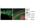
A: Immunofluorescence staining of rat neuron-glial cultures with R-1687-100 (red). The rabbit antibody shows strong and specific staining of neuronal cilia. Neuronal alpha-II spectrin was double-labelled with M-1575-100 (green), and cell nuclei were visualized with a DNA dye (blue).
B: Immunohistochemistry of mouse brain sections with R-1687-100 (green). Brains were fixed by transcardial perfusion with 4% paraformaldehyde and stained with R-1687-100 antibody (green) and anti-Fox3/NeuN monoclonal antibody M-377-100 antibody (red). R-1687-100 specifically labels cilia next to the pyramidal neurons in CA1 hippocampus region, but not in other part of the brain. Cell nuclei were stained with DAPI (blue).
C: Western blot analysis of rat olfactory epithelium (Lane 1) and frontal cortex (Lane 2). R-1687-100 reveals protein bands at about 200 kDa in olfactory epithelium, a tissue which is rich in cilia. The frontal cortex contains less cilia, and the protein is also less heavily glycosylated, so that a much less prominent band is seen at about 160 kDa.
Click on image to zoom
B: Immunohistochemistry of mouse brain sections with R-1687-100 (green). Brains were fixed by transcardial perfusion with 4% paraformaldehyde and stained with R-1687-100 antibody (green) and anti-Fox3/NeuN monoclonal antibody M-377-100 antibody (red). R-1687-100 specifically labels cilia next to the pyramidal neurons in CA1 hippocampus region, but not in other part of the brain. Cell nuclei were stained with DAPI (blue).
C: Western blot analysis of rat olfactory epithelium (Lane 1) and frontal cortex (Lane 2). R-1687-100 reveals protein bands at about 200 kDa in olfactory epithelium, a tissue which is rich in cilia. The frontal cortex contains less cilia, and the protein is also less heavily glycosylated, so that a much less prominent band is seen at about 160 kDa.
Ships: 1-2 business days