Anti-Actin Antibody (5J11)
Our Anti-Actin mouse monoclonal primary antibody detects bovine, horse, human, pig, and rat Actin, and is IgG. It is validated for use in FC, ICC, WB.
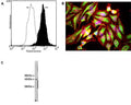
![Left: Actin expression in HeLa cells visualized with mouse antibody to actin (green) by Immunocytochemistry. Vimentin was co-stained with chicken antibody to vimentin (C-1409-50, red). Blue: DAPI nuclear stain.The actin antibody labels the submembranous actin-rich cytoskeleton, stress fibers and bundles of actin associated with cell adhesion sites. The vimentin antibody stains a different cytoskeletal network, the intermediate or 10 nm filaments. Right: Western blot analysis of tissue and cell lysates probed with mouse anti-actin antibody (red, lanes 2-7). [1] protein standard, [2] rat brain, [3] mouse brain, [4] NIH-3T3, [5] HEK293, [6] HeLa, [7] SH-SY5Y cells. The same blot was simultaneously probed chicken antibody against UCHL1 (C-1406-50, green), a marker of neuronal lineage cells.](http://www.antibodiesinc.com/cdn/shop/files/m-1646-100-ihc-wb_120x120.jpg?v=1759273309)

A: Specific staining of Actin expressed in human neuroblastoma SH-SY5Y cell line by Flow Cytometry using cat # M-1646-100. Fixing and Permeabilization of cells: Absolute methanol (10 minutes in ice) and 0.1% Tween-20 in PBS, Blocking: 1% BSA, Primary antibody: M-1646-100, 2 μg per ~10^6 cells, 30 minutes at room temperature, Secondary antibody: Goat anti-mouse PE (1:100), 20 minutes in dark at room temperature. Negative control: Non-specific Control IgG, clone X63 (cat # M-1249-200, black dashed). Data and results were generated using Orflo MoxiflowTM instrument and protocols. B: HeLa cells stained with M-1646-100 (red) and also with our chicken polyclonal antibody to Vimentin (C-1409-50). The actin antibody stains the submembranous actin rich cytoskeleton and also stress fibers, bundles of actin associated with adhesion sites. The vimentin antibody stains a quite different cytoskeletal network, the intermediate filaments. The blue stain reveals DNA in the nuclei of these cells. C: Crude extract of HeLa cells. The antibody recognizes the ~42 kDa protein.
Click on image to zoom
Ships: 1-2 business days