Anti-Microtubule-associated protein Tau (MAPT) Antibody
Our Anti-Microtubule-associated protein Tau (MAPT) rabbit polyclonal primary antibody detects human and pig Microtubule-associated protein Tau (MAPT), and is whole serum. It is validated for use in ELISA, WB.

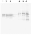
Western blot analysis on recombinant human Tau purified from E.Coli (lanes 1-3; 10, 20 and 40 µg respectively) and on porcine brain lysate (lanes 4-6; 20, 40, 80 ng respectively) under reducing conditions using Rabbit antibody to human Microtubule-associate protein Tau: whole serum (R-184-100) at a dilution of 1: 500. This antibody detected recombinant human Tau with the smaller fragments likely representing degradation products from the purified protein, which has been expressed in E. Coli. In the porcine cytosol, this antibody detected several bands that represents the different Tau isoforms and likely phosphorylated species. This polyclonal antibody is independent of phosphorylation.
Click on image to zoom
SKU: R-184-100
Product Details
Microtubule-associated protein Tau (MAPT)
FUNCTION: Promotes microtubule assembly and stability, and might be involved in the establishment and maintenance of neuronal polarity. The C-terminus binds axonal microtubules while the N-terminus binds neural plasma membrane components, suggesting that tau functions as a linker protein between both. Axonal polarity is predetermined by tau localization (in the neuronal cell) in the domain of the cell body defined by the centrosome. The short isoforms allow plasticity of the cytoskeleton whereas the longer isoforms may preferentially play a role in its stabilization. SUBCELLULAR LOCATION: Cytoplasm; cytosol. Cell membrane. Mostly found in the axons of neurons, in the cytosol and in association with plasma membrane components. ALTERNATIVE PRODUCTS: 8 named isoforms produced by alternative splicing. Additional isoforms seem to exist. Isoforms differ from each other by the presence or absence of up to 5 of the 15 exons. One of these optional exons contains the additional tau/MAP repeat. TISSUE SPECIFICITY: Expressed in neurons. Isoform PNS-tau is expressed in the peripheral nervous system while the others are expressed in the central nervous system. DEVELOPMENTAL STAGE: Four-repeat (type II) tau is expressed in an adult-specific manner and is not found in fetal brain, whereas three-repeat (type I) tau is found in both adult and fetal brain. DOMAIN: The tau/MAP repeat binds to tubulin. In Alzheimer disease, the neuronal cytoskeleton in the brain is progressively disrupted and replaced by tangles of paired helical filaments and straight filaments, mainly composed of hyperphosphorylated forms of Microtubule-associated protein Tau. Defects in Microtubule-associated protein Tau are a cause of frontotemporal dementia and parkinsonism linked to chromosome 17, as well as a number of other neurodegenerative diseases.
Whole serum
Polyclonal
Mixed
ELISA, WB
Rabbit
Recombinant human Microtubule-associated protein Tau, purified from E.coli.
Human
Human, Pig
Spin vial briefly before opening. Reconstitute in 100 µL sterile-filtered, ultrapure water. Centrifuge to remove any insoluble material. After reconstitution keep aliquots at -20°C for higher stability, and at 2-8°C with an appropriate antibacterial agent. Glycerol (1:1) may be added for an additional stability. Avoid repetitive freeze/thaw cycles.
Lyophilized
WB: 1:500
This antibody has been shown to detect the purified recombinant Tau expressed in E.Coli as well as a number of Tau isoforms in porcine cytosol.
Unconjugated
Specificity was demonstrated by Western blot. 20 ng recombinant Tau is easily detected with smaller fragments likely representing degradation products from the purified protein, which has been expressed in E. Coli. This antiserum is known to react with human and pig Microtubule-associated Tau proteins. Other species have not yet been tested.
For research use only.
United States
12 months after date of receipt (unopened vial).
Neurofibrillary tangle protein; Paired helical filament-tau; PHF-tau; MAPT; MTBT1; TAU
25°C (ambient)