Anti-Microtubule-associated proteins 1A/1B light chain 3A (MAP1LC3A) Antibody (BS405)
Our Anti-Microtubule-associated proteins 1A/1B light chain 3A (MAP1LC3A) mouse monoclonal primary antibody detects human, mouse (predicted), and rat Microtubule-associated proteins 1A/1B light chain 3A (MAP1LC3A), and is IgG. It is validated for use in FC, ICC, WB.
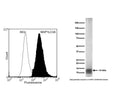
Left: Flow cytometry analysis of human SHSY-5Y cells of MAP1LC3A with M-1725-100. Fixing and Permeabilization of cells: Absolute methanol and 0.1% Tween-20 in PBS, Primary antibody: Mouse Monoclonal antibody to MAP1LC3A (cat # M-1725-100, 2 μg per ~10^6 cells) for 15 minutes at room temperature, Secondary antibody: Goat anti-mouse PE labelled secondary antibody (1:1000 fold dilution) with incubation for 15 minutes in dark at room temperature. Non-specific Control IgG, clone X63 (cat # M-1249-200) was used under same conditions as negative control (black dashed). Flow cytometry data and results were generated using Orflo Moxiflow instrument and protocols. The data demonstrates specific staining of MAP1LC3A expressed in human neuronal cell line SHSY-5Y using cat # M-1725-100.
Right: Detection of MAP1LC3A in human SHSY-5Y cell lysates (20 µg/lane) by Western Blotting. SDS-PAGE: 4-20% Bis-Tris denatured, reduced; Transfer: Tris-Glycine buffer; Membrane: nitrocellulose (0.2 µm); Blocking: 5% skim milk in TBST, 1 hour at RT; Primary antibody: overnight at 4°C (10 µg/mL); Secondary antibody: anti-mouse-HRP (1/10000) 2 hours at RT; Detection: Enhanced Chemiluminescence Substrate with LiCor C-DiGit Blot Scanner. The broad band seen on WB indicates binding of Cat # M-1725-100 to a broad protein band with a Molecular Weight of ~14 kDa. Predicted molecular weight of MAP1LC3A is ~14.5 kDa.
Click on image to zoom
Right: Detection of MAP1LC3A in human SHSY-5Y cell lysates (20 µg/lane) by Western Blotting. SDS-PAGE: 4-20% Bis-Tris denatured, reduced; Transfer: Tris-Glycine buffer; Membrane: nitrocellulose (0.2 µm); Blocking: 5% skim milk in TBST, 1 hour at RT; Primary antibody: overnight at 4°C (10 µg/mL); Secondary antibody: anti-mouse-HRP (1/10000) 2 hours at RT; Detection: Enhanced Chemiluminescence Substrate with LiCor C-DiGit Blot Scanner. The broad band seen on WB indicates binding of Cat # M-1725-100 to a broad protein band with a Molecular Weight of ~14 kDa. Predicted molecular weight of MAP1LC3A is ~14.5 kDa.
SKU: M-1725-100
Product Details
Microtubule-associated proteins 1A/1B light chain 3A (MAP1LC3A)
MAP1A and MAP1B are microtubule-associated protein which mediate the physical interactions between microtubules and components of the cytoskeleton (probably involved in autophagosome formation). MAP1A and MAP1B each consist of a heavy chain subunit and 3 different light chain subunits (LC1, LC2 and LC3). MAP1LC3A is one of the light chain subunits and can associate with either MAP1A or MAP1B. The precursor form of MAP1LC3A is cleaved by APG4/ATG4B to form the cytosolic form LC3-1. This is activated by APG7L/ATG7, transferred to ATG3 and conjugated to phospholipid to form the membrane-bound form, LC3-II. MAP1LC3A is most abundant in heart, brain, liver, skeletal muscle and testis but is absent in thymus and peripheral leukocytes.
IgG
Monoclonal
BS405
Flow, ICC, WB
Mouse
14 kDa
A synthetic peptide (C-RSFADRCKEVQQI) corresponding to amino acids 11-23 of human MAP1LC3 A protein was conjugated to a carrier protein and the complex used as the immunogen. This human sequence is homologous with mouse and rat MAP1LC3 A.
Human
Human, Mouse, Rat
Spin vial briefly before opening. Reconstitute in 100 µL sterile-filtered, ultrapure water. Centrifuge to remove any insoluble material. Final buffer contains no preservatives.After reconstitution divide into aliquots and store at -20°C for a higher stability. Antibody contains no preservatives. Store at 2-8°C with an appropriate antibacterial agent. Use sterile methods. Highest purity Glycerol (1:1) may be added for an additional stability when stored at refrigerated or freezing temperatures. Avoid repetitive freeze/thaw cycles.
Lyophilized
Protein G purified mouse immunoglobulin
Lyophilized from PBS, pH 7.4, containing 3% trehalose without preservatives.
WB: 10 µg/mL
ICC: 1-2 µg/mL
ICC: 1-2 µg/mL
Flow cytometry: 20 µg/mL
Unconjugated
WB confirmed binding of the antibody to a broad protein band with a Molecular Weight of ~14 kDa. Rat. The antibody is expected to react with mouse MAP1LC3A protein due to 100% sequence homology.
For research use only.
United States
12 months after date of receipt (unopened vial).
Microtubule-associated proteins 1A/1B light chain 3A; MAP1A/MAP1B LC3 A; MAP1A/1B light chain 3 A; MAP1 light chain 3-like protein 1; Microtubule-associated protein 1 light chain 3 alpha; Autophagy-related protein LC3 A; Autophagy-related ubiquitin-like modifier LC3 A; APG8a; MAP1LC3A
25°C (ambient)

