Anti-Lysosomal Associated Membrane Protein 1 (LAMP1) Antibody (LAMP1)
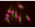
Our Anti-Lysosomal associated membrane protein 1 (LAMP1) mouse monoclonal primary antibody detects human Lysosomal associated membrane protein 1 (LAMP1), and is IgG. It is validated for use in ICC, WB.
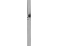
SKU: M-1652-100
Product Details
Lysosomal associated membrane protein 1 (LAMP1)
LAMP1 (Lysosomal Associated Membrane Protein 1, also known as CD107a, lysosomal associated membrane glycoprotein 1, LGP120 and LAMPA) is a protein primarily associated with the lysosomal membrane. In a typical cell LAMP1 is associated with spherical vesicles located next to the nucleus and the microtubule organizing center (1). LAMP1 is found on the cell surface of lymphocytes undergoing degranulation, a process in which cytoplasmic vesicles fuse with the plasma membrane, and this phenomena resulted in discovery of LAMP1 as a CD protein. The LAMP1 protein has a large N-terminal region which is inside the lysosome, hence topologically external to the cell, which is often referred to as the lumenal domain (2). The lumenal domain consists of two homologous globular segments separated by a proline rich sequence. Next there is a single membrane spanning domain and a short 11 amino acid C-terminal cytoplasmic tail. This tail region contains, at the extreme C-terminus, a so-called YXXI motif which is responsible for the sorting of the intact molecule to the endosome and lysozome, where Y = tyrosine, I = isoleucine and X = almost any amino acid (3). This motif is found in several other lysosomal proteins, where it functions in the same way. There are 417 amino acids in the human LAMP1 molecule, giving a native molecular weight of 44.8 kDa. However the N-terminal lumenal segment of LAMP1 is very heavily and variably glycosylated due to the presence of 18 N-linked glycosylation sites, so that on SDS-PAGE and on Western blots the protein runs as a diffuse band at 90-120 kDa. Antibodies to LAMP1 are therefore excellent markers of lysosomes in mammalian cells, though some LAMP1 may also be seen on late endosomes and on the plasma membrane.
IgG
Monoclonal
LAMP1
IgG1
ICC, WB
Mouse
90-120 kDa
Recombinant LAMP1 expressed and purified from E. coli.
Human
Human
Spin vial briefly before opening. Reconstitute with 100 µL sterile-filtered, ultrapure water to achieve a 1 mg/mL concentration. Centrifuge to remove any insoluble material. After reconstitution of lyophilized antibody, aliquot and store at -20°C for a higher stability. Avoid freeze-thaw cycles.
Lyophilized
Protein G purified
Lyophilized from PBS buffer pH 7.2-7.6 with 0.1% trehalose, and sodium azide
WB: 1:5000-1:10000
Unconjugated
The antibody reacts with a diffuse band at ~90 kDa to 120 kDa by Western blot on HeLa cell extract. It has also been used successfully for immunocytochemistry.
For research use only.
United States
12 months after date of receipt (unopened vial).
Lysosomal Associated Membrane Protein 1, also known as CD107a, lysosomal associated membrane glycoprotein 1, LGP120 and LAMPA
25°C (ambient)

