Anti-Growth Associated Protein 43 (GAP-43) Antibody (GAP43)
Our Anti-Growth associated protein 43 (GAP-43) mouse monoclonal primary antibody detects mouse and rat Growth associated protein 43 (GAP-43), and is IgG. It is validated for use in FC, ICC, WB.
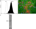
A: Flow Cytometry analysis of GAP43 expressed in human neuroblastoma SH-SY5Y cell line. Fixing and permeabilization of cells: Absolute methanol (10 minutes in ice) and 0.1% Tween-20 in PBS; Blocking: 1% BSA; Primary antibody: Mouse Monoclonal antibody to GAP43 (cat # M-1650-100, 2 μg per ~106 cells) for 30 minutes at room temperature; Secondary antibody: Goat anti-mouse PE (1:100 dilution), incubation for 20 minutes in dark at room temperature. Non-specific Control IgG, clone X63 (cat # M-1249-200) was used as negative control under same conditions. Data and results were generated using Orflo MoxiflowTM instrument and protocols. B: Immunofluorescence analysis of mixed neuron-glial cultures stained with mouse anti-GAP43 (green) and rabbit anti-MAP2 (red). Blue: DNA staining. The GAP43 antibody stains the plasma membrane of neurons and is particularly concentrated in dendrites. C: Western Blot analysis of GAP43 expression in whole rat spinal cord lysates. The antibody recognizes the ~43 kDa protein.
Click on image to zoom
SKU: M-1650-100
Product Details
Growth associated protein 43 (GAP-43)
GAP43 is very abundant protein which is found concentrated in neurons. One group discovered it as one of three proteins which becomes unregulated during the regeneration of the toad optic nerve (1). Three GAPs (Growth associated proteins) were discovered, and the number 43 comes from the apparent SDS-PAGE molecular weight of the one named GAP43. The HGNC name for this protein is, not surprisingly, GAP43. Later work showed that GAP43 does not run on SDS-PAGE in a fashion which accurately reflects its molecular weight, and that GAP43 proteins from different species may run at different apparent molecular weights. Partly due to these features GAP43 were independently discovered by several different groups and therefore has several alternate names, such as protein F1, pp46, neuromodulin, neural phosphoprotein B-50 and calmodulin-binding protein P-57. In each case the number reflects the apparent SDS-PAGE molecular weight, and underlines the unusual properties of this molecule. Mammalian GAP43 proteins contains only 226-243 amino acids, and so the real molecular weight is 23.61-25.14 kDa. GAP43 has been extensively studied and is known to be a major protein kinase C substrate and to bind calmodulin avidly. GAP43 is anchored to the plasma membrane by palmitoylation modifications.
IgG
Monoclonal
GAP43
IgG1
Flow, ICC, WB
Mouse
43 kDa
C-terminal peptide of rat and mouse GAP43, which is KEDPEADQEHA, to which an N terminal Cysteine residue was added to allow chemical coupling to Keyhole Limpet Hemocyanin carrier protein.
Rat
Mouse, Rat
Spin vial briefly before opening. Reconstitute with 100 µL sterile-filtered, ultrapure water to achieve a 1 mg/mL concentration. Centrifuge to remove any insoluble material. After reconstitution of lyophilized antibody, aliquot and store at -20°C for a higher stability. Avoid freeze-thaw cycles.
Lyophilized
Affinity Purified
Lyophilized from PBS buffer pH 7.2-7.6 with 0.1% trehalose, and sodium azide
WB: 1:5000-1:10000
ICC: 1:1000-1:5000
ICC: 1:1000-1:5000
Use 2 ug/10^6 cells for Flow cytometry.
Unconjugated
The antibody reacts with a 43 kDa band by Western blot on rat spinal cord lysate. It has also been used successfully for immunocytochemistry.
For research use only.
United States
12 months after date of receipt (unopened vial).
protein F1, neuromodulin, neural phosphoprotein B-50, axonal membrane protein GAP-43, calmodulin-binding protein P-57
25°C (ambient)

