Anti-Neurofilament light polypeptide (NF-L), DG-Sensor™ Antibody
Our Anti-Neurofilament light polypeptide (NF-L) [degenerated] rabbit polyclonal primary antibody detects bovine, human, mouse, pig, and rat Neurofilament light polypeptide (NF-L) [degenerated], and is IgG. It is validated for use in ELISA, IF, ICC, IHC, WB.
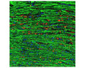

SKU: R-2124-100
Product Details
ICC: 1:1000
The antibody recognizes NF-L in reduced westerns regardless of the disease state.
Block and permeabilize sections in 5-10% normal goat serum or serum of the species the secondary antibodies were made in, in PBS plus 1% Triton-100 (PBST) for 1 hour with slight agitation, followed by primary antibody incubations and fluorescent secondary identification. High primary antibody dilutions require refrigerated, overnight incubations for best results. Recommended fixation is 4% PFA fixed, frozen tissue 20-50 microns; other fixation methods have not been tested and are not recommended at this time.
Degeneration-specific detection is fixation and antigen recovery dependent. This antibody reacts to a peptide epitope within amino acids 311-375 of the human NF-L. This peptide epitope is uncovered in degenerating cells but not normal cells. However, the specific reactivity of this epitope is sensitive to tissue treatments and could become exposed in healthy cells under some conditions. For example, treatment of the fixed tissue with high temperatures, proteinase, or other denaturants may cause the reactive epitope to become exposed in healthy cells, leading to a false positive. Biosensis recommends experimenting with treated and untreated tissues when first using these antibodies if degeneration specificity is desired. The exact conditions and dilutions must be determined experimentally by the end user.
This antibody will detect NL-L protein in paraffin-embedded tissues; however, the degeneration-specific detection can be problematic in paraffin tissues, particularly if Heat-Induced Epitope Retrieval (HIER), or other common antigen recovery methods are used. This is because the reactive epitope, which is covered in healthy cells but exposed in degenerative cells, could become accessible to the antibody in healthy cells, leading to false positives. For this reason, paraffin-embedded tissues are not recommended if degeneration-specific detection is desired.