Anti-Microtubule-associated protein tau (MAPT) Antibody
Our Anti-Microtubule-associated protein tau (MAPT) chicken polyclonal primary antibody detects bovine (predicted), chicken (predicted), horse (predicted), human, mouse (predicted), pig (predicted), and rat (predicted) Microtubule-associated protein tau (MAPT), and is IgY preparation. It is validated for use in ICC, WB.
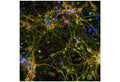
Immunofluorescence analysis of tau protein expression in cortical neuron-glial culture from E20 rat. The chicken anti-tau antibody (green, 1:2,000) stains perikarya, dendrites and axons of neurons, while a anti-MAP2 antibody labels only dendrites and perikarya (red). Perikarya and dendrites appear orange-yellow, since they contain both MAP2 and tau proteins. Blue: DAPI nuclear stain.
Click on image to zoom
Bovine, Chicken, Horse, Human, Mouse, Pig, Rat
ICC, WB
Chicken
SKU: C-1691-100
Product Details
Microtubule-associated protein tau (MAPT)
FUNCTION: Promotes microtubule assembly and stability, and might be involved in the establishment and maintenance of neuronal polarity. The C-terminus binds axonal microtubules while the N-terminus binds neural plasma membrane components, suggesting that tau functions as a linker protein between both. Axonal polarity is predetermined by tau localization (in the neuronal cell) in the domain of the cell body defined by the centrosome. The short isoforms allow plasticity of the cytoskeleton whereas the longer isoforms may preferentially play a role in its stabilization. SUBCELLULAR LOCATION: Cytoplasm; cytosol. Cell membrane. Mostly found in the axons of neurons, in the cytosol and in association with plasma membrane components. ALTERNATIVE PRODUCTS: 8 named isoforms produced by alternative splicing. Additional isoforms seem to exist. Isoforms differ from each other by the presence or absence of up to 5 of the 15 exons. One of these optional exons contains the additional tau/MAP repeat. TISSUE SPECIFICITY: Expressed in neurons. Isoform PNS-tau is expressed in the peripheral nervous system while the others are expressed in the central nervous system. DEVELOPMENTAL STAGE: Four-repeat (type II) tau is expressed in an adult-specific manner and is not found in fetal brain, whereas three-repeat (type I) tau is found in both adult and fetal brain. DOMAIN: The tau/MAP repeat binds to tubulin. In Alzheimer disease, the neuronal cytoskeleton in the brain is progressively disrupted and replaced by tangles of paired helical filaments and straight filaments, mainly composed of hyperphosphorylated forms of Microtubule-associated protein Tau. Defects in Microtubule-associated protein Tau are a cause of frontotemporal dementia and parkinsonism linked to chromosome 17, as well as a number of other neurodegenerative diseases.
IgY preparation
Polyclonal
IgY
ICC, WB
Chicken
48 kDa to 67 kDa, with an additional band at 100 kDa
Recombinant full length version of the shortest human tau isoform purified from E. coli.
Human
Bovine, Chicken, Horse, Human, Mouse, Pig, Rat
Spin vial briefly before opening. Reconstitute with 100 µL sterile-filtered, ultrapure water. Centrifuge to remove any insoluble material. Store lyophilized, unopened vial at 2-8°C or lower. After reconstitution, prepare aliquots and store at -20°C to -80°C for a higher stability. Avoid freeze-thaw cycles.
Lyophilized
Lyophilized IgY preparation, with sodium azide.
WB: 1:5000-1:10000
ICC: 1:500-1:1000
ICC: 1:500-1:1000
Unconjugated
The antibody reacts with multiple closely spaced bands covering the region of the blot from 48 kDa to 67 kDa, with an additional band at 100 kDa. It has also been used successfully for immunocytochemistry. Expected to react with horse, cow, pig, chicken, rat and mouse.
For research use only.
United States
12 months after date of receipt (unopened vial).
Neurofibrillary tangle protein; Paired helical filament-tau; PHF-tau; MAPT; MTBT1; TAU;
25°C (ambient)

