Anti-Microtubule-associated protein 2 (MAP2) Antibody
Our Anti-Microtubule-associated protein 2 (MAP2) chicken polyclonal primary antibody detects bovine, human, mouse, and rat Microtubule-associated protein 2 (MAP2), and is IgY preparation. It is validated for use in ICC, IHC-Frozen, IHC-Paraffin-embedded, WB.
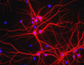
![Left: Analysis of MAP2 expression in cortical neuron-glial cell culture from E20 rat by Immunocytochemistry with chicken anti-MAP2 (1:10,000, red), co-stained with a mouse antibody to MAP-tau (green). Blue: DAPI nuclear stain. The MAP2 antibody stains dendrites and perikarya of neurons, while the MAP-tau antibody labels neuronal perikarya, dendrites and also axonal process. As a result perikarya and dendrites appears orange-yellow, since they contain both MAP2 and tau, while axons are green. Right: Western blot analysis of whole brain tissue lysates using chicken antibody to MAP2 (1:50,000). [1] protein standard, [2] adult rat brain, [3] embryonic E20 rat brain, [4] adult mouse brain. A strong band at around 280 kDa corresponds to two major isoforms of MAP2 protein referred to as MAP2A and MAP2B. Smaller fragments of these isoforms are also detected if the antibody is used at lower dilutions.](http://www.antibodiesinc.com/cdn/shop/files/c-1382-50-ihc-wb_120x120.jpg?v=1759273993)

View of mixed neuron/glial cultures stained with Chicken polyclonal antibody to Microtubule Associated Protein 2 C-1382-50 (red). The perikarya and dendrites of neurons are strongly and specifically stained with this antibody while the axons of the neurons and the processes of all other cell types in these cultures (astrocytes, oligodendrocytes, microglia, endothelia and fibroblasts) are all negative. Cell nuclei are visualized with DAPI DNA stain.
Click on image to zoom