Anti-mCherry Antibody
Our Anti-mCherry chicken polyclonal primary antibody detects mCherry, and is concentrated purified IgY. It is validated for use in ICC, WB.
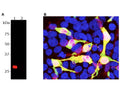
![Left: HEK293 cells transfected with a mCherry (red) were stained with chicken antibody to mCherry (green). Blue: DAPI nuclear stain. Transfected cells appear bright red, and co-stain for mCherry chicken antibody. Superimposition of both signals results in yellow-orange colour. Right: Western blot analysis of transfected and control HEK293 cell lysates, and recombinant proteins with chicken antibody to mCherry (1:2,000, green). [1] protein standard, [2] HEK293 control, [3] HEK293 cells transfected with mCherry, [4] recombinant mCherry protein, [5] recombinant GFP protein, [6] HEK293 transfected with GFP. The major band at about 30 kDa corresponds to mCherry protein and the slightly larger recombinant form runs at about 33 kDa due to presence of a His tag and other vector derived sequences. No cross-reactivity with GFP protein is observed. The same blot was simultaneously probed with a mouse antibody to HSP60 (red), which reveals a band at 60 kDa only in cell lysates.](http://www.antibodiesinc.com/cdn/shop/files/C-1655-100-ihc-wb_120x120.jpg?v=1759274145)

A: Western blot analysis of HEK293 cells transfected with pFin-EF1-mCherry vector. C-1655-100 (1:2,000 dilution) reveals a strong protein band in transfected cells (Lane 1). Non-transfected control cells (Lane 2) show no mCherry protein band.
B: Immunofluorescence analysis of mCherry-transfected HEK293 cells with C-1655-100. Specificity of C-1655-100 for mCherry is shown by superimposing mCherry fluorescence (red) with C-1655-100 antibody staining (green), which results in a yellow color. mCherry staining is also seen in the nucleus, which might be due to degradation of mCherry molecules, causing the release of the low molecular weight mCherry fluorochrome.
Click on image to zoom
B: Immunofluorescence analysis of mCherry-transfected HEK293 cells with C-1655-100. Specificity of C-1655-100 for mCherry is shown by superimposing mCherry fluorescence (red) with C-1655-100 antibody staining (green), which results in a yellow color. mCherry staining is also seen in the nucleus, which might be due to degradation of mCherry molecules, causing the release of the low molecular weight mCherry fluorochrome.