Anti-Capsaicin receptor (VR1/TrpV1) Antibody (BS397)
Our Anti-Capsaicin receptor (TrpV1) mouse monoclonal primary antibody detects guinea pig (predicted), mouse, and rat Capsaicin receptor (TrpV1), and is IgG. It is validated for use in FC, WB.
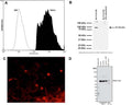
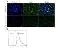

A: Analysis of TRPV1 expression in rat PC12 cell line by Flow Cytometry. Fixing and permeabilization of cells: Absolute methanol (10 minutes in ice) and 0.1% Tween-20 in PBS, Blocking: 1% BSA, Primary antibody: Mouse Monoclonal antibody to TRPV1 (cat # M-1714-100, 2 µg per ~10^6 cells) for 30 minutes at RT, Secondary antibody: Goat anti-mouse PE labeled secondary antibody (1:100 dilution), 20 minutes in dark at room temperature. Negative control: Non-specific Control IgG, clone X63 (cat # M-1249-200, black dashed). Data and results were generated using Orflo MoxiflowTM instrument and protocols.
B: Western blot of TrpV1 in rat PC12 cell lysates (80 µg/lane). M-1714-100 detects TrpV1 protein at 95-100 kDa. SDS-PAGE: denatured and reduced; Transfer: Tris-Glycine buffer; Membrane: nitrocellulose (0.45 µm); Blocking: 5% skim milk in TBST, 1 hour at RT; Primary antibody: overnight at 4°C (2 µg/mL); Secondary antibody: anti-mouse-HRP (1/6000) 2 hours at RT; Detection: Chemiluminiscence.
C: Immunohistochemical staining of TrpV1 in mouse dorsal root ganglia. Immunoreactivity was visualized with anti-mouse-Cy3 conjugate (red). Magnification: 20x. Courtesy P. Vilimas, Flinders University Adelaide.
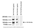
D: Western blot (denatured and reduced) of TrpV1 in cell lysates of forskolin and NGF stimulated 50B11 hybrid mouse x rat DRG cell lines and NGF-stimulated PC12 cells (10 µg/lane). M-1714-100 detects monomeric TrpV1 protein at 95-100 kDa. Primary antibody: 1 µg/mL (4°C overnight). Detection: Chemiluminiscence. Courtesy Dr. D. Matusica, Flinders University.
Click on image to zoom
B: Western blot of TrpV1 in rat PC12 cell lysates (80 µg/lane). M-1714-100 detects TrpV1 protein at 95-100 kDa. SDS-PAGE: denatured and reduced; Transfer: Tris-Glycine buffer; Membrane: nitrocellulose (0.45 µm); Blocking: 5% skim milk in TBST, 1 hour at RT; Primary antibody: overnight at 4°C (2 µg/mL); Secondary antibody: anti-mouse-HRP (1/6000) 2 hours at RT; Detection: Chemiluminiscence.
C: Immunohistochemical staining of TrpV1 in mouse dorsal root ganglia. Immunoreactivity was visualized with anti-mouse-Cy3 conjugate (red). Magnification: 20x. Courtesy P. Vilimas, Flinders University Adelaide.
D: Western blot (denatured and reduced) of TrpV1 in cell lysates of forskolin and NGF stimulated 50B11 hybrid mouse x rat DRG cell lines and NGF-stimulated PC12 cells (10 µg/lane). M-1714-100 detects monomeric TrpV1 protein at 95-100 kDa. Primary antibody: 1 µg/mL (4°C overnight). Detection: Chemiluminiscence. Courtesy Dr. D. Matusica, Flinders University.