Anti-Calretinin-binding protein (CR) Antibody (3G9)
Our Anti-Calretinin-binding protein (CR) mouse monoclonal primary antibody detects bovine, human, mouse, and rat Calretinin-binding protein (CR), and is affinity-purified. It is validated for use in IHC-Frozen, WB.
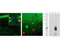
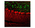

Left and Middle: Detection of calretinin immunoreactivity (red) in adult mouse brain hippocampal sections (Left) and adult rat cortical sections (Middle) by Immunohistochemistry. The calretinin antibody (1:1,000) stains a small number of interneurons in the stratum radiatum of CA1 region (Left), while Fox/NeuN (R-3770-100, green) is expressed in most neurons in the brain. As a result, co-labelled neurons that stain positive for calretinin appear yellow. In rat cortex (Right), calretinin (red) is expressed in a small population of interneurons concentrated in Layer 4 area, while calbindin (C-1798-50, green) is expressed in cells concentrated in Layer 2/3. Because each antibody specifically labels a different population of cells exclusively, the cells are either stained green or red in the cortex. Blue: DAPI nuclear stain. Insets are high-magnification images of the boxed area in each picture. IHC method: 45 um sections, tissue fixed by transcardial perfusion with 4% paraformaldehyde. Right: Western blot analysis of calretinin. The antibody binds strongly and cleanly to a calretinin band at ~32 kDa in cow cerebellum homogenate (Lane 1). Specificity for calretinin and absecnce of cross-reactivity with other related calcium-binding proteins is shown by probing recombinant proteins: secretagogin (Lane 2), parvalbumin (Lane 3), calretinin (Lane 4), calbindin (Lane 5).
Click on image to zoom