Anti-Cav1.2 Ca2+ Channel Antibody (N263/31)

Our Anti-Cav1.2 Ca2+ channel mouse monoclonal primary antibody from NeuroMab is produced in-house from hybridoma clone N263/31. It detects human, mouse, and rat Cav1.2 Ca2+ channel, and is purified by Protein A chromatography. It is great for use in ICC, WB.
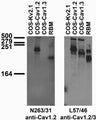

Tissue and transfected cell immunoblot: extracts of rat brain membrane (RBM) and COS cells transiently transfected with untagged Cav1.2, Cav1.3 or Kv2.1 plasmids and probed with N263/31 (left) and L57/46 (right) TC supe.
Click on image to zoom
![]()
Guinea Pig, Human, Mouse, Rabbit, Rat
ELISA, ICC, IHC, WB
Mouse
Ships: 1-2 business days
Product Specific References for Applications and Species
- FRET: Rat
- Immunocytochemistry: Guinea Pig | Mouse | Rat
- Immunohistochemistry: Human | Mouse
- Immunoprecipitation: Rat
- PLA: Human | Mouse
- STED: Rabbit
- Western Blot: Guinea Pig | Human | Mouse | Rat
| Fluorescence Resonance Energy Transfer: Rat | ||
| PMID | Dilution | Publication |
| 27076616 | 1:100 | Bannister, J.P., et al. 2016. Rab25 influences functional Cav1.2 channel surface expression in arterial smooth muscle cells. American Journal of Physiology. Cell Physiology, C885-C895. |
| Immunocytochemistry: Guinea Pig | ||
| PMID | Dilution | Publication |
| 31403402 | 1:200 | Morgenstern, T.J., et al. 2019. A potent voltage-gated calcium channel inhibitor engineered from a nanobody targeted to auxiliary CaVβ subunits. Elife, e49253. |
| Immunocytochemistry: Mouse | ||
| PMID | Dilution | Publication |
| 37702787 | 1:100 | Akin, EJ, et al. 2023. ANO1, CaV1.2, and IP3R form a localized unit of EC-coupling in mouse pulmonary arterial smooth muscle. The Journal of General Physiology, 0. |
| 37438479 | 1:100 | Cserne Szappanos, H., et al. 2023. Cytoskeletal Disarray Increases Arrhythmogenic Vulnerability During Sympathetic Stimulation in a Model of Hypertrophic Cardiomyopathy. Scientific Reports, 11296. |
| 34750263 | 1:5 (supe) | Vierra, N.C., et al. 2021. Regulation of neuronal excitation-transcription coupling by Kv2.1-induced clustering of somatic L-type Ca2+ channels at ER-PM junctions. PNAS: USA, . |
| 33082339 | 10ug/ml | Prada, M.P., et al. 2020. AKAP5 complex facilitates purinergic modulation of vascular L-type Ca2+ channel CaV1.2. Nature Communications, 5303. |
| 31663850 | 1:5 (supe) | Vierra, N.C., et al. 2019. Kv2. 1 mediates spatial and functional coupling of L-type calcium channels and ryanodine receptors in mammalian neurons. Elife, e49953. |
| Immunocytochemistry: Rat | ||
| PMID | Dilution | Publication |
| 34431981 | 1:100 | Isensee, J., et al. 2021. Depolarization induces nociceptor sensitization by CaV1.2-mediated PKA-II activation. Journal of Cell Biology, . |
| 27364017 | not listed | Eichel, C.A., et al. 2016. Lateral Membrane-Specific MAGUK CASK Down-Regulates NaV1.5 Channel in Cardiac Myocytes. Circulation Research, 544-56. |
| Immunohistochemistry: Human | ||
| PMID | Dilution | Publication |
| 28119464 | 1:1000 | Nystoriak, M.A., et al. 2017. Ser1928 phosphorylation by PKA stimulates the L-type Ca2+ channel CaV1.2 and vasoconstriction during acute hyperglycemia and diabetes. Scientific Signaling, eaaf9647. |
| Immunohistochemistry: Mouse | ||
| PMID | Dilution | Publication |
| 37507375 | 5ug/ml | Casas, M., et al. 2023. NPC1-dependent alterations in KV2.1–CaV1.2 nanodomains drive neuronal death in models of Niemann-Pick Type C disease. Nature Communications, 4553. |
| 34750263 | 1:5 (supe) | Vierra, N.C., et al. 2021. Regulation of neuronal excitation-transcription coupling by Kv2.1-induced clustering of somatic L-type Ca2+ channels at ER-PM junctions. PNAS: USA, . |
| Immunoprecipitation: Rat | ||
| PMID | Dilution | Publication |
| 24681347 | 1:500 | Liu, W., et al. 2014. KCNE2 modulates cardiac L-type Ca(2+) channel.. Journal of Molecular Cellular Cardiology, 208-218. |
| PLA: Human | ||
| PMID | Dilution | Publication |
| 28119464 | 1:1000 | Nystoriak, M.A., et al. 2017. Ser1928 phosphorylation by PKA stimulates the L-type Ca2+ channel CaV1.2 and vasoconstriction during acute hyperglycemia and diabetes. Scientific Signaling, eaaf9647. |
| PLA: Mouse | ||
| PMID | Dilution | Publication |
| 38860414 | not listed | Xu, B, et al. 2024. Differential Downregulation of β1-Adrenergic Receptor Signaling in the Heart. Journal of the American Heart Association, e033733. |
| STED: Rabbit | ||
| PMID | Dilution | Publication |
| 29549309 | not listed | Zhang, X.D., et al. 2018. Coupling of SK channels, L-type Ca2+ channels, and ryanodine receptors in cardiomyocytes. Scientific Reports, 4670. |
| Western Blot: Guinea Pig | ||
| PMID | Dilution | Publication |
| 26764482 | 1:500 | Nassal, D.M., et al. 2016. Myocardial KChIP2 expression in guinea pig resolves an expanded electrophysiologic role. PLoS One, e0146561. |
| Western Blot: Human | ||
| PMID | Dilution | Publication |
| 24401693 | 1:200 | Duan, L., et al. 2014. Stem cell derived basal forebrain cholinergic neurons from Alzheimer''s disease patients are more susceptible to cell death.. Molecular Neurdegeneration, 3. |
| 23858011 | 1:250 | Narayanan, D., et al. 2013. Smooth muscle cell transient receptor potential polycystin-2 (TRPP2) channels contribute to the myogenic response in cerebral arteries.. Journal of Physiology, 5031-5046. |
| Western Blot: Mouse | ||
| PMID | Dilution | Publication |
| 32442021 | not listed | Gupte, R., et al. 2020. Glucose-6-phosphate dehydrogenase increases Ca2+ currents by interacting with Cav1.2 and reducing intrinsic inactivation of the L-type calcium channel. American Journal of Physiology. Heart and Circulatory Physiology, H144-H158. |
| Western Blot: Rat | ||
| PMID | Dilution | Publication |
| 33453240 | 1:1000 | Nasu, F., et al. 2021. Azelnidipine treatment reduces the expression of Cav1.2 protein. Life Sciences, 119043. |
| 31337710 | not listed | Jiang, M., et al. 2019. S-Palmitoylation of junctophilin-2 is critical for its role in tethering the sarcoplasmic reticulum to the plasma membrane. Journal of Biological Chemistry, 13487-13501. |
| 25610372 | 1:1000 | Perissinotti, P.P., et al. 2015. Calcium current homeostasis and synaptic deficits in hippocampal neurons from Kelch-like 1 knockout mice.. Frontiers in Cellular Neuroscience, 444. |
| 24703904 | 1:1000 | Perissinotti, P.P., et al. 2014. Down-regulation of endogenous KLHL1 decreases voltage-gated calcium current density.. Cell Calcium, 269-280. |
| 23858011 | 1:250 | Narayanan, D., et al. 2013. Smooth muscle cell transient receptor potential polycystin-2 (TRPP2) channels contribute to the myogenic response in cerebral arteries.. Journal of Physiology, 5031-5046. |
| 23568894 | not listed | Bannister, J.P., et al. 2013. The voltage-dependent L-type Ca2+ (CaV1.2) channel C-terminus fragment is a bi-modal vasodilator. Journal of Physiology, 2987-2998. |