Anti-Gephyrin Antibody (L106/83)

Our Anti-Gephyrin mouse monoclonal primary antibody from NeuroMab is produced in-house from hybridoma clone L106/83. It detects human, mouse, and rat Gephyrin, and is purified by Protein A chromatography. It is great for use in AT, IHC, ICC, WB.
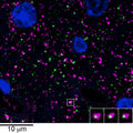

Array tomography immunofluorescence of an LRWhite-embedded 70 nm section from adult mouse cortex with L106/83 (green), rabbit mAb GAD2 (Cell Signaling 5843, magenta) and DAPI (blue). The insert shows three consecutive sections through the synapse that is marked with a white box. Image courtesy of Kristina Micheva (Stanford).
Click on image to zoom
![]()
Human, Mouse, Rat
AT, ELISA, ICC, IHC, WB
Mouse
Ships: 1-2 business days
Product Specific References for Applications and Species
| Immunohistochemistry: Mouse | ||
| PMID | Dilution | Publication |
| 34576197 | 1:100 | Kuljis, D.A., et al. 2021. Gephyrin-Lacking PV synapses on neocortical pyramidal neurons. International Journal of Molecular Science, 10032. |
| 33704414 | 1:100 | Micheva, K.D., et al. 2021. Conduction velocity along the local axons of parvalbumin interneurons correlates with the degree of axonal myelination. Cerebral Cortex, 3374-3392. |
| 31554841 | 1:100 | Simhal, A.K., et al. 2019. Multifaceted changes in synaptic composition and astrocytic involvement in a mouse model of fragile X syndrome. Scientific Reports, 13855. |
| Western Blot: Mouse | ||
| PMID | Dilution | Publication |
| 37759527 | not listed | Gruol, DL, et al. 2023. Impact of Elevated Brain IL-6 in Transgenic Mice on the Behavioral and Neurochemical Consequences of Chronic Alcohol Exposure. Cells, 0. |
| 36775097 | not listed | Gruol, D.L., et al. 2023. Neuroimmune Interactions with Binge Alcohol Drinking in the Cerebellum of IL-6 Transgenic Mice. Neuropharmacology, 109455. |
| 35789851 | 1:1000 | Huo, Y., et al. 2022. Prkn Knockout Mice Show Autistic-like Behaviors and Aberrant Synapse Formation. iScience, 104573. |
| 32634532 | not listed | Gruol, D.L., et al. 2020. Alcohol and IL-6 Alter Expression of Synaptic Proteins in Cerebellum of Transgenic Mice With Increased Astrocyte Expression of IL-6. Neuroscience, 124-137. |
| 32202499 | 1:5000 | Zhang, R.S., et al. 2020. Latrophilin-2 and latrophilin-3 are redundantly essential for parallel-fiber synapse function in cerebellum. Elife, e54443. |
| 29787738 | not listed | Gruol, D.L., et al. 2018. Altered Brain Activity During Withdrawal from Chronic Alcohol is Associated with Changes in IL-6 Signal Transduction and GABAergic Mechanisms in Transgenic Mice with Increased Astrocyte Expression of IL-6. Neuropharmacology., 32-46. |
| 29787738 | not listed | Gruol, D.L., et al. 2018. Altered brain activity during withdrawal from chronic alcohol is associated with changes in IL-6 signal transduction and GABAergic mechanisms in transgenic mice with increased astrocyte expression of IL-6. Neuropharmacology, 32-46. |