Anti-Gephyrin Antibody (L106/4)

Our Anti-Gephyrin mouse monoclonal primary antibody from NeuroMab is produced in-house from hybridoma clone L106/4. It detects human, mouse, and rat Gephyrin, and is purified by Protein A chromatography. It is great for use in AT, IHC, WB.
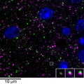

array tomography immunofluorescence of an LRWhite-embedded 70 nm section from adult mouse cortex with L106/4 (green), rabbit mAb GAD2 (Cell Signaling 5843, magenta) and DAPI (blue). The insert shows three consecutive sections through the synapse that is marked with a white box. Image courtesy of Kristina Micheva (Stanford).
Click on image to zoom
Ships: 1-2 business days
Product Specific References for Applications and Species
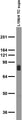
- Western Blot: Mouse
| Western Blot: Mouse | ||
| PMID | Dilution | Publication |
| 39388350 | 1:1000 | Dunham, TL, et al. 2024. WWC2 modulates GABAA-receptor-mediated synaptic transmission, revealing class-specific mechanisms of synapse regulation by WWC family proteins. Cell Reports, 114841. |
| 38507236 | 1:2000 | McCormick, LE, et al. 2024. The E3 ubiquitin ligase TRIM9 regulates synaptic function and actin dynamics in response to netrin-1. Molecular Biology of the Cell, mbcE23120476. |