Anti-Ubiquitin carboxyl-terminal hydrolase isozyme L1 (UCHL1) Antibody (BH7)
Our Anti-Ubiquitin carboxyl-terminal hydrolase isozyme L1 (UCHL1) mouse monoclonal primary antibody detects bovine, human, mouse, pig, and rat Ubiquitin carboxyl-terminal hydrolase isozyme L1 (UCHL1), and is IgG. It is validated for use in ICC, IHC-Frozen, WB.
![An image showing a section of rat spinal cord stained with Mouse monoclonal antibody to Ubiquitin C Terminal Hydrolase 1 [BH7] M-1407-100 (red) and Rabbit polyclonal antibody to neurofilament H R-1388-50 (green). The large cells are a-motorneurons and Ubiquitin C Terminal Hydrolase 1 (UCHL1) fills the cytoplasm of their perikarya and dendrites. The neurofilament NF-H antibody binds primarily to phosphorylated axonal forms of NF-H, and so stains axons coursing between the large motor neurons.](http://www.antibodiesinc.com/cdn/shop/files/m-1407-100-ihc-1_120x120.jpg?v=1759273248)
![Left: Analysis of UCHL1 expression in rat hippocampal section by Immunohistochemistry. UCHL1 protein was visualized with mouse antibody to UCHL1 (green, 1:5,000), and section co-stained with rabbit anti-FOX3/NeuN (R-3770-100, red, 1:2,000). Blue: DAPI nuclear stain. IHC Method: Following transcardial perfusion of rat with 4% paraformaldehyde, brain was post-fixed for 24 hours, cut to 45 um, and free-floating sections were stained. The UCHL1 antibody stains the cell body and dendrites of hippocampal neurons, while the FOX3 antibody labels nuclei of the neuronal cells. Right: Western blot analysis of UCHL1 expression in tissue lysates (green, 1:10,000). [1] protein standard, [2] rat brain, [3] rat spinal cord, [4] mouse brain, [5] mouse spinal cord, [6] pig brain, [7] pig spinal cord. The single band at 24 kDa corresponds to the UCHL1 protein.](http://www.antibodiesinc.com/cdn/shop/files/m-1407-100-ihc-wb_120x120.jpg?v=1759273248)
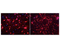
![An image showing a section of rat spinal cord stained with Mouse monoclonal antibody to Ubiquitin C Terminal Hydrolase 1 [BH7] M-1407-100 (red) and Rabbit polyclonal antibody to neurofilament H R-1388-50 (green). The large cells are a-motorneurons and Ubiquitin C Terminal Hydrolase 1 (UCHL1) fills the cytoplasm of their perikarya and dendrites. The neurofilament NF-H antibody binds primarily to phosphorylated axonal forms of NF-H, and so stains axons coursing between the large motor neurons.](http://www.antibodiesinc.com/cdn/shop/files/m-1407-100-ihc-1_1600x.jpg?v=1759273248)
An image showing a section of rat spinal cord stained with Mouse monoclonal antibody to Ubiquitin C Terminal Hydrolase 1 [BH7] M-1407-100 (red) and Rabbit polyclonal antibody to neurofilament H R-1388-50 (green). The large cells are a-motorneurons and Ubiquitin C Terminal Hydrolase 1 (UCHL1) fills the cytoplasm of their perikarya and dendrites. The neurofilament NF-H antibody binds primarily to phosphorylated axonal forms of NF-H, and so stains axons coursing between the large motor neurons.
Click on image to zoom