Anti-Tyrosine Hydroxylase (TH) Antibody
Our Anti-Tyrosine Hydroxylase (TH) rabbit polyclonal primary antibody detects guinea pig, mouse, and rat Tyrosine Hydroxylase (TH), and is whole serum. It is validated for use in IHC-Frozen, WB.


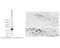
Left: Western Blot analysis of tyrosine hydroxylase (TH) expression in PC12 cell lysate (6 ug protein/lane). The anti-TH rabbit antibody (1:300) detects one specific band at ~60 kDa corresponding to the expected molecular weight of TH. Method: SDS Page: denaturing and reducing, 4-12% Bis-Tris gel; Transfer: Tris-Glycine (Towbin's buffer) with 20% methanol; Membrane: PVDF (0.45 um); Blocking: 5% skim milk in TBST, 1 hr at RT; Primary antibody: R-118-100 (1:300), overnight at 2-8°C; Secondary antibody: donkey anti-rabbit (1:25,000), 1 hr at RT; Detection: Chemiluminescence. Right: Detection of TH-immunoreactivity in dopaminergic neurons in the rat zona incerta in formalin-fixed floated cryostat section by Immunohistochemistry. TH was visualized with the rabbit polyclonal antiserum (R-118-100; 1:100,000) using the biotinylated secondary antibody-ABC method and nickel-diaminobenzidine chromogen. Photo courtesy of Dr. Erik Hrabovszky, Hungarian Academy of Sciences, Budapest, Hungary.
Click on image to zoom
SKU: R-118-100
Product Details
Tyrosine Hydroxylase (TH)
Tyrosine hydroxylase (TH) is the rate-limiting enzyme in the synthesis of the catecholamines dopamine, epinephrine and norepinephrine. Therefore the regulation of the TH enzyme represents the central means for controlling the synthesis of these important catecholamines. FUNCTION: Plays an important role in the physiology of adrenergic neurons. CATALYTIC ACTIVITY: L-tyrosine + tetrahydrobiopterin + O2 = 3,4-dihydroxy-L-phenylalanine + 4a-hydroxytetrahydrobiopterin. COFACTOR: Fe(2+) ion. ENZYME REGULATION: Phosphorylation leads to an increase in the catalytic activity. PATHWAY: Catecholamine biosynthesis; first step. SUBUNIT: Homotetramer. PTM: In vitro, phosphorylation of Ser-19 increases the rate of Ser-40 phosphorylation, which results in enzyme opening and activation. SIMILARITY: Belongs to the biopterin-dependent aromatic amino acid hydroxylase family. The presence of different DNA sequences at the TH locus confers susceptibility to various disorders of the brain including manic-depression and schizophrenia. Parkinson's disease is also considered a TH deficiency as low dopamine levels are a consistent neurochemical abnormality.
Whole serum
Polyclonal
Mixed
IHC, WB
Rabbit
60 kDa
A synthetic peptide (PRFIGRRQSLIEDARK) as part of human Tyrosine Hydroxylase (63-78) conjugated to KLH has been used as the immunogen. The peptide is homologous with the corresponding sequence derived from TH protein in rat (31-47).
Rat
Guinea Pig, Mouse, Pig, Rat
Spin vial briefly before opening. Reconstitute in 100 µL sterile-filtered, ultrapure water. Centrifuge to remove any insoluble material. After reconstitution keep aliquots at -20°C for a higher stability, and at 2-8°C with an appropriate antibacterial agent. Glycerol (1:1) may be added for an additional stability. Avoid repetitive freeze/thaw cycles.
Lyophilized
WB: 1:100-1:500
IHC: 1:2000-1:5000
IHC: 1:2000-1:5000
This antibody is for detection of tyrosine hydroxylase (TH) containing neurons exhibiting an intense labelling with a negligible background. This antiserum has proven useful for staining of catecholaminergic neurons. It stains dendritic processes and fine nerve terminals. We recommend mouse or rat brain containing catecholaminergic neurons as a positive control for this antibody, for example brain stem or striatum.
Western blotting (WB): Antibody has been tested on RIPA-extracted PC12 cell lysate and shown to be specific for TH (~60 kDa). Tissue homogenates show a higher level of non-specific binding and presence of uncharacterized bands. Affinity-purified anti-TH antibody (R-148-50) is recommended for tissue homogenates.
Unconjugated
IHC on brain shows a pattern of staining specific for TH containing neurons. This antibody is known to react with rat, mouse and guinea pig. Cross reactivity with other species has not yet been tested.
For research use only.
United States
12 months after date of receipt (unopened vial).
TH; Tyrosine hydroxylase; Tyrosine 3-monooxygenase; L-tyrosine hydroxylase; Tyrosine 3-hydroxylase
25°C (ambient)