Anti-TAR DNA-binding protein 43 (TDP-43) Antibody (3H8)
Our Anti-TAR DNA-binding protein 43 (TDP-43) mouse monoclonal primary antibody detects human, mouse, other mammals (predicted), and rat TAR DNA-binding protein 43 (TDP-43), and is IgG. It is validated for use in ICC, IHC-Frozen, WB.
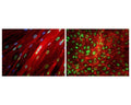
![Mouse monoclonal antibody to TAR DNA-binding protein 43 [3H8] M-1403-100 was used to stain a section of formalin fixed adult rat brain, specifically the hippocampus. Hippocampal neuron nuclei are stained strongly. Chicken polyclonal antibody to GFAP C-1373-50 (green) shows the processes of astrocytic glial cells. Nuclei of all cells are revealed with DAPI DNA stain (blue). The TAR DNA-binding protein 43 antibody stains neuronal nuclei strongly and the nuclei of some non-neuronal cells much more weakly. Neuronal nuclei therefore look crimson, since they are both red due to the content of TAR DNA-binding protein 43 and blue due to their content of DNA, stained blue with DAPI.](http://www.antibodiesinc.com/cdn/shop/files/m-1403-100-ihc-1_120x120.jpg?v=1759273235)
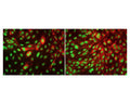
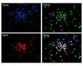
![Left: Analysis of TDP43 expression in rat hippocampus section by Immunohistochemistry. Section was stained with mouse anti-TDP43 antibody (red, 1:2,000), and co-stained with chicken antibody to GFAP (C-1373-50, green, 1:5,000). Blue: DAPI nuclear stain. IHC method: Following transcardial perfusion of rat with 4% paraformaldehyde, brain was post-fixed for 24 hours, cut to 45 um sections, and free-floating sections were stained. The TDP43 protein is concentrated in the nuclei of hippocampal neurons, while the GFAP antibody stains the intermediate filament network of astroglial cells. Right: Western blot analysis of whole brain lysates and nuclear extracts for TDP43 expression (green, 1:2,000). [1] protein standard, [2] rat brain, [3] rat brain nuclear extract, [4] mouse brain, [5] mouse brain nuclear extract. A strong band at 43 kDa corresponds to TDP43 protein.](http://www.antibodiesinc.com/cdn/shop/files/m-1403-100-ihc-wb_120x120.jpg?v=1759273236)
![Mouse monoclonal antibody to TAR DNA-binding protein 43 [3H8] M-1403-100 was used to stain a section of formalin fixed adult rat brain, specifically the hippocampus. Hippocampal neuron nuclei are stained strongly. Chicken polyclonal antibody to GFAP C-1373-50 (green) shows the processes of astrocytic glial cells. Nuclei of all cells are revealed with DAPI DNA stain (blue). The TAR DNA-binding protein 43 antibody stains neuronal nuclei strongly and the nuclei of some non-neuronal cells much more weakly. Neuronal nuclei therefore look crimson, since they are both red due to the content of TAR DNA-binding protein 43 and blue due to their content of DNA, stained blue with DAPI.](http://www.antibodiesinc.com/cdn/shop/files/m-1403-100-ihc-1_1600x.jpg?v=1759273235)
Mouse monoclonal antibody to TAR DNA-binding protein 43 [3H8] M-1403-100 was used to stain a section of formalin fixed adult rat brain, specifically the hippocampus. Hippocampal neuron nuclei are stained strongly. Chicken polyclonal antibody to GFAP C-1373-50 (green) shows the processes of astrocytic glial cells. Nuclei of all cells are revealed with DAPI DNA stain (blue). The TAR DNA-binding protein 43 antibody stains neuronal nuclei strongly and the nuclei of some non-neuronal cells much more weakly. Neuronal nuclei therefore look crimson, since they are both red due to the content of TAR DNA-binding protein 43 and blue due to their content of DNA, stained blue with DAPI.
Click on image to zoom