Magic Red® Fluorescent Cathepsin L Assay Kit

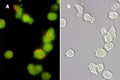
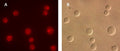
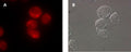
Quantitatively monitor intracellular cathepsin-L activity over time in vitro. The Magic Red® substrate fluoresces red upon cleavage by active cathepsin enzymes. The signal can be analyzed using fluorescence microscopy or a fluorescence plate reader.


SKU: 941

Size: 25 Tests
Price:
Sale price$206.00

Our Magic Red® Cathepsin assay kits enable researchers to quantitate and monitor intracellular cathepsin B, K, or L activity over time in vitro. The Magic Red® (MR) reagent is a non-cytotoxic substrate that fluoresces red upon cleavage by active cathepsin enzymes.

Elevated cathepsin enzyme activity in serum or the extracellular matrix often signifies a number of gross pathological conditions. Cathepsin-mediated diseases include: Alzheimer’s; numerous types of cancer; autoimmune related diseases like arthritis; and the accelerated breakdown of bone structure seen with osteoporosis. Up-regulated cathepsin B and L activity has been linked to several types of cancer. These include cancer of the colon, pancreas, ovaries, breast, lung, and skin (melanoma). Upregulation of cathepsin K has been shown in lung tumors. Increased cathepsin K activity has also been linked to degenerative bone diseases including osteopetrosis and post-menopausal osteoporosis.

Cathepsins are usually characterized as members of the lysosomal cysteine protease (active site) family and the cathepsin family name has been synonymous with lysosomal proteolytic enzymes. In actuality, the cathepsin family also contains members of the serine protease (cathepsin A and G) and aspartic protease (cathepsin D and E) families as well. These enzymes exist in their processed form as disulfide-linked heavy and light chain subunits with molecular weights ranging from 20-35 kDa. Cathepsin C is the noted exception, existing as an oligomeric enzyme with a MW ~200 kDa. Initially synthesized as inactive zymogens, cathepsins are post-translationally processed into their active configurations after passing through the endoplasmic reticulum and subsequent incorporation into the acidic environment of the lysosomes.

Magic Red® detection substrates utilize the photostable red fluorophore, cresyl violet. When bi-substituted via amide linkage to two cathepsin target peptide sequences, such as (leucine-arginine)2, the bi-substituted cresyl violet is non-fluorescent. Following enzymatic cleavage at one or both arginine (R) amide linkage sites, the mono and non-substituted cresyl violet fluorophores generate red fluorescence when excited at 550-590 nm. Our Magic Red® cathepsin B substrate, MR-(RR)2, is comprised of cresyl violet coupled to two pairs of the amino acid sequence, arginine-arginine (RR), which is the preferential target sequence for cathepsin B. In our cathepsin K substrate, MR-(LR)2, cresyl violet is coupled to two pairs of leucine- arginine (LR). Our MR cathepsin L substrate, MR-(FR)2, contains two pairs of phenylalanine-arginine (FR) coupled to cresyl violet. Cathepsins, like most other crucial cell survival enzymes, are somewhat permissive in the target amino acid sequence they will recognize and cleave. Although Magic Red® substrates contain the amino acid target sequence preferred by a particular cathepsin enzyme, they can also recognize other active cathepsins or proteases when they are present. We encourage validation of cathepsin activity by an orthogonal technique.

To use Magic Red®, add the substrate directly to the cell culture media, incubate, and analyze. Because MR is cell-permeant, it easily penetrates the cell membrane and the membranes of the internal cellular organelles - no lysis or permeabilization steps are required. If cathepsin enzymes are active, they will cleave off the two dipeptide cathepsin targeting sequences and allow the cresyl violet fluorophore to become fluorescent upon excitation. The red fluorescent product will stay inside the cell and will often aggregate inside lysosomes (cathepsins are lysosomal) and other areas of low pH, such as inside the mitochondria. As protease activity progresses and more MR substrate is cleaved, the signal will intensify as the red fluorescent product accumulates within various organelles, enabling researchers to watch the color develop over time and quantify cathepsin B, K, or L activity. By varying the duration and concentration of exposure to the MR substrate, a picture can be obtained of the relative abundance of cathepsin enzymatic activity. Positive cells will fluoresce red and have pronounced red lysosomes and mitochondria. Negative cells will exhibit very low levels of background red fluorescence evenly distributed throughout the cell. This background level of substrate activity could be the result of constitutively synthesized serine proteases that target analogous amino acid sequences for hydrolysis. Please note that Magic Red® substrates can undergo spontaneous hydrolysis over time, resulting in increased background fluorescence. Appropriate controls are necessary for accurate interpretation of the results. There is no interference from pro-cathepsin forms of the enzymes. If the treatment or experimental condition stimulates cathepsin activity, cells containing elevated levels of cathepsin activity will appear brighter red than cells with lower levels of cathepsin activity.

The MR fluorophore, cresyl violet, fluoresces red when excited at 550-590 nm. The red fluorescent signal can be monitored with a fluorescence microscope or plate reader. It has an optimal excitation of 592 nm and emission of 628 nm. Hoechst 33342 is included with the kit to concurrently label nuclei after labeling with MR. It is revealed under a microscope using a UV-filter with excitation at 365 nm and emission at 480 nm. Acridine orange (AO) is also included in the kit to identify lysosomes and other intracellular organelles. It is revealed under a microscope using excitation at 480 nm and emission at >540 nm, or alternatively with excitation at 550 nm and emission at >610 nm.

MR-FR2
Cathepsin L
592 nm / 628 nm
Fluorescence Plate Reader, Fluorescence Microscope
Cell culture, tissue
2-8°C
Domestic: Overnight Delivery; International: Priority Shipping
United States
  1. Prepare samples and controls
  2. Reconstitute Magic Red by adding 50 µL DMSO.
  3. Dilute Magic Red 1:10 by adding 450 µL diH2O.
  4. Add 20 µL Magic Red to each sample (~ 500 µL aliquot of cultured cells).
  5. Incubate while protected from light.
  6. Watch color start to develop within 15 minutes.
  7. If desired, label with additional stains, such as Hoechst, DAPI, Acridine orange, or an antibody.
  8. If desired, fix or embed cells.
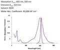
  9. Analyze with a fluorescence microscope or fluorescence plate reader. Magic Red has an optimal excitation at 592 nm and emission at 628 nm.

If working with adherent cells, please see the manual for additional protocols.

Kit 941: 25 Tests
  • Magic Red Reagent (MR-FR2), 1 25-test vial, #6137
  • Hoechst 33342 Stain, 1 mL, #639
  • Acridine Orange Stain, 0.5 mL, #6130
  • Kit Manual
  • Kit 942: 100 Tests
  • Magic Red Reagent (MR-FR2), 1 100-test vial, #6138
  • Hoechst 33342 Stain, 1 mL, #639
  • Acridine Orange Stain, 0.5 mL, #6130
  • Kit Manual
  • Product Specific References

    PMID Publication
    38515365Sung, H.K., et al. 2024. Ischemia-induced cardiac dysfunction is exacerbated in adiponectin-knockout mice due to impaired autophagy flux. Clinical and translational science, e13758.
    37883971Ebner, M., et al. 2023. Nutrient-regulated control of lysosome function by signaling lipid conversion. Cell, .
    37142604Zeng, J., et al. 2023. Restoration of lysosomal acidification rescues autophagy and metabolic dysfunction in non-alcoholic fatty liver disease. Nature communications, 2573.
    37375535Zhang, C., et al. 2023. Bis-Benzylisoquinoline Alkaloids Inhibit Porcine Epidemic Diarrhea Virus by Disrupting Virus Entry. Pathogens (Basel, Switzerland).
    37417221Ezz, M.A., et al. 2023. Cathepsin L regulates oocyte meiosis and preimplantation embryo development. Cell proliferation, e13526.
    35535798Rodrigues, P.M., et al. 2022. LAMP2 regulates autophagy in the thymic epithelium and thymic stroma-dependent CD4 T cell development. Autophagy, 44940.
    35977928Ratto, E., et al. 2022. Direct control of lysosomal catabolic activity by mTORC1 through regulation of V-ATPase assembly. Nature communications, 4848.
    36535215Wu, K., et al. 2022. The endo-lysosomal regulatory protein BLOC1S1 modulates hepatic lysosomal content and lysosomal lipolysis. Biochemical and biophysical research communications, 44936.
    33372271Yang, Y., et al. 2021. Porphyromonas gingivalis facilitated the foam cell formation via lysosomal integral membrane protein 2 (LIMP2). Journal of periodontal research.
    33629936Wu, K., et al. 2021. BLOC1S1/GCN5L1/BORCS1 is a critical mediator for the initiation of autolysosomal tubulation. Autophagy.

    Question: When using a fluorescence plate reader staining for adherent cell, do I have to detach the cell by trypsin treatment? In that case, Is there anything to be aware of?

    Answer: If you are using adherent cells, you do not necessarily need to detach them. If your plate reader is capable of reading from the bottom, you can grow your adherent culture in a tissue culture plate with black walls and a clear bottom.

    Bulk Order Magic Red® Fluorescent Cathepsin L Assay Kit

    Related Products

    Recently Viewed