Poly Caspase SR-VAD-FMK Kit
This kit uses the red fluorescent probe SR-VAD-FMK to label active caspases within living cells. Caspase positive cells may be distinguished from caspase negative cells by fluorescence microscopy or fluorescence plate reader. HTS adaptable (microplate).

Apoptosis is an evolutionarily conserved form of cell suicide, which follows a specialized cellular process. The central component of this process is a cascade of proteolytic enzymes called caspases. These enzymes participate in a series of reactions that are triggered in response to pro-apoptotic signals and result in cleavage of protein substrates, causing the disassembly of the cell. Caspases have been identified in organisms ranging from C. elegans to humans. The mammalian caspases play distinct roles in apoptosis and inflammation. In apoptosis, caspases are responsible for proteolytic cleavages that lead to cell disassembly (effector caspases), and are involved in upstream regulatory events (initiator caspases). An active caspase consists of two large (~20 kD) and two small (~10 kD) subunits to form two heterodimers which associate in a tetramer. As is common with other proteases, caspases are synthesized as precursors that undergo proteolytic maturation, either autocatalytically or in a cascade by enzymes with similar specificity. Caspase enzymes specifically recognize a 4 amino acid sequence (on their substrate) which necessarily includes an aspartic acid residue. This residue is the target for the cleavage reaction, which occurs at the carbonyl end of the aspartic acid residue. Caspases can be detected via immunoprecipitation, immunoblotting techniques using caspase specific antibodies, or by employing fluorogenic substrates which become fluorescent upon cleavage by the caspase.
Our Poly Caspase SR-VAD-FMK Kit uses a novel approach to detect active caspases . The methodology is based on sulforhodamine labeled fluoromethyl ketone (FMK)-peptide inhibitors of caspases. These inhibitors are cell permeable and non-cytotoxic. Once inside the cell, the inhibitor binds covalently to the active caspase . Cells that contain bound inhibitor can be analyzed by fluorescence microscopy or fluorescence plate reader.
SR100-2: 100 Tests
Product Specific References
| PMID | Publication |
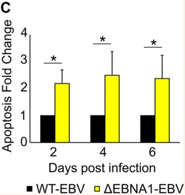
| 34781735 | Westhoff Smith, D., et al. 2021. The Epstein-Barr Virus Oncogene EBNA1 Suppresses Natural Killer Cell Responses and Apoptosis Early after Infection of Peripheral B Cells. mBio, e0224321. |
| 21088132 | Vereide, D. T., et al. 2011. Lymphomas differ in their dependence on Epstein-Barr virus. Blood, 1977-85. |